Development of the Ear


Development of the Ear


Michael John Wareing

Anil K. Lalwani

Abbas A. Anwar

Robert K. Jackler


The development of the structures necessary for transmission of sound information from the environment to the auditory cortex is a complex and interwoven process. An understanding of the major developmental steps and their interrelations is desirable because therein lies the key to understanding many conditions encountered by otolaryngologists. Awareness of the developmental process alerts the surgeon to anatomic associations and explains important departures from normal.

Abnormal development is important for its clinical effects, but it also has a role in unraveling the complexities of normal development. The critical period of ear development begins in the third week after fertilization, the inner ear appearing first. The inner, middle, and outer portions of the ear have different embryologic origins, and development can be arrested at any stage. The result is a range of abnormalities from mild to severe. In view of the different origins, a disorder in one part does not necessarily signify a disorder in another, but proximity in terms of time of development, originating tissue, anatomic characteristics, and function does mean that multiple malformations are possible. The disorders can be caused by an inborn genetic error, either inherited or spontaneous, or by a teratogenic influence during organogenesis. The tissues of the head and neck are derived from all three layers of the embryo—ectoderm, mesoderm, and endoderm. The neural crest cells play a special role in the head and neck, where they constitute most of the skeletal and connective tissue. These cells arise from the ectodermal layer at the junction where the neural tube begins to fold. All divisions of the ear contain some neural crest tissue. The mesodermal proportion in the head and neck is less than in the rest of the body.

The story of ear development goes back to the time life itself was in its infancy. Fish seem to be the first hearing organisms, with development of a hearing organ from an internal balance organ. Even at this early evolutionary stage, the hair-cell design now so widespread was in use.

Both amphibians and reptiles inherited the balance labyrinth of fish but went on to develop auditory labyrinths of their own, having branched from the line of fish before acquisition of a hearing organ. The need to hear in air resulted in development of a conductive apparatus to correct the impedance mismatch of sound arriving in air but having to be transmitted into the liquid of the labyrinth. Mammalian design continued from the basic reptilian design with, in particular, the addition of rows of hair cells, an independent cochlear nerve, changes in the middle-ear conduction system, and protective external auditory canals (1). Throughout this chapter, we separate development of the ear into its component parts as an aid to understanding. It is important, however, to remember that these changes occurred in a simultaneous manner. An overview of ear development is presented in Table 140.1.


AURICULAR DEVELOPMENT

In keeping with its recent evolutionary appearance, the auricle of the external ear begins its development later than do other components of the ear. From the fifth week of gestation, three hillocks arise on the first branchial (mandibular) arch (hillocks 1, 2, and 3) and three arise on the second branchial (hyoid) arch (hillocks 4, 5, and 6) on either side of the first branchial cleft (Fig. 140.1). Hillocks 1 and 6 are the first to be identifiable separately, but by the sixth week, all are distinct. The lobule also can be identified on the second arch. By the eighth week, the auricle has an identifiable structure, and the contributions of the hillocks to the adult form can be recognized: hillock 1, tragus; hillock 2, crus helicis; hillock 3, ascending helix; hillock 4, horizontal helix, upper portion of scapha, and antihelix; hillock 5, descending helix, middle portion of the scapha, and antihelix; and hillock 6, antitragus and inferior aspect of the helix (2). Although this is the majority view, there is uncertainty about the origin of the crus helicis and
ascending helix; some believe that these structures can arise from the second arch (3). By approximately 18 weeks’ gestation, the auricle has achieved essentially adult form, although it continues to grow in childhood with changes continuing into late adult life.


TABLE 140.1 OVERVIEW OF EAR DEVELOPMENT


Fetal Age (wk)


Outer Ear


Middle Ear


Inner Ear


3


Otic placode develops. VC ganglia appear


4


EAC begins


Tubotympanic recess is apparent


Otocyst present


5


Hillocks become evident


Ossicles begin to condense in mesenchyme


Otocyst begins to divide into vestibular and cochlear areas


Semicircular canals begin to outpouch; VC ganglion divides


6


All hillocks are distinct


Malleus and incus are identifiable as cartilaginous models


Superior SCC completed. Utricle and saccule are present; cochlear duct begins


7


Maculae present; sensory ridges in the cochlea appear


8


Auricle has identifiable structure; deep meatus is apparent as epithelial strand


Incudomalleolar and incudostapedial joints form


Ductus reuniens is identifiable; 1.5 cochlear turns; cristae present


Vacuoles develop in vascular precartilage surrounding membranous labyrinth; cartilaginous model of otic capsule forms


9


Tympanic membrane has trilaminar structure


Nerve fibers enter sensory epithelium; oval window develops


10


Stapes loses annular form; facial nerve runs through middle ear


2.5 cochlear turns


11


Hair cells present in cochlea; synaptic connections are present


12


Tympanic ring begins to ossify


Otoconial membrane is present; cochlear duct changes to triangular shape


16


Malleus, incus, and stapes begin to ossify


Ossification of the otic capsule begins


18


Auricle has adult form


20


Meatal plug begins to disintegrate


Tympanic cavity begins to open


Cochlear duct reaches full length; membranous labyrinth is full size


22


Antrum begins to develop


Tunnel of Corti present at all levels; basal turn of cochlea is functional


23


Otic capsule ossification is complete


24


Perilymphatic space is completed


26


Facial nerve makes second genu in adult position


28


EAC is fully open


30


Malleus and incus are ossified


34


Ossicles lie within open middle ear space


Mastoid air cells begin to develop


VC, vestibulocochlear; SCC, semicircular canal; EAC, external auditory canal.


DEVELOPMENTAL ANOMALIES

A wide spectrum of pinna deformity exists, from anotia, in which there is no development, to a small but normally formed pinna. Microtia encapsulates the wide spectrum between anotia and normality. The superior portion of the auricle usually is severely malformed or absent. The presence of a deformity of the pinna can indicate further defects of the auditory system. Although this is less common with some minor deformities, severe cases of microtia and anotia are almost always associated with atresia of the external auditory canal and defects of the middle ear (see later).

The etiology of auricular abnormalities remains unclear, but evidence has implicated both environmental and genetic factors. Environmental risk factors for microtia are numerous and include anemia, advanced maternal or paternal age, male gender, race, and multiple births. In addition, mothers with chronic type I diabetes are at significantly higher risk for having children with microtia (4). Microtia may also have genetic risk factors, as autosomal recessive and dominant forms of the deformity
with variable expression and incomplete penetrance have been reported. More recently, specific genes, such as the

Gsc homeobox gene and the BMP5 maternal peptide gene, have been implicated as possible predisposing genes for microtia (5). Auricular abnormalities also are present in all the common chromosomal abnormalities, including trisomy 13, 18, and 22 as well as chromosomal translocations and microdeletions, and thus, are useful markers of these conditions. Classification of deformities of the pinna and treatment are discussed in Chapters 190 and 191.


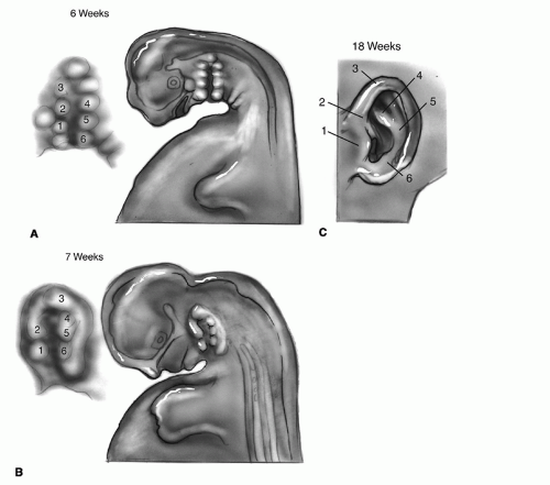
Figure 140.1 Development of the auricle. A: Six hillocks form on the first and second branchial arches. All can be identified at 6 weeks’ gestation. B: Seven-week stage. C: By 18 weeks, the adult form is recognizable.


DEVELOPMENT OF THE EXTERNAL AND MIDDLE EAR


External Auditory Canal

The external auditory canal begins to form in the fourth week of gestation (Fig. 140.2). The first branchial cleft, between the first and second branchial arches, widens, and the ectoderm proliferates to form a pit, which comes into apposition with the endoderm of the first pharyngeal pouch. This pit is the forerunner of the cartilaginous external auditory canal. This arrangement is temporary because mesenchymal growth separates the cleft and the pouch. The deep portion of the external auditory canal is apparent from the eighth week of gestation as a strand of epithelial cells running down to the disc-shaped precursor of the tympanic membrane (3). At approximately 28 weeks’ gestation, this epithelial core has canalized from the medial to the lateral aspect to allow communication with the tympanic membrane. The epithelial core is the precursor of the bony external auditory canal.


Tympanic Membrane

The tympanic membrane has a trilaminar origin of ectoderm from the floor of the first branchial cleft laterally as the epidermal layer, endoderm of the first pharyngeal pouch medially as the mucosal layer, and neural crest mesenchyme with cephalic mesoderm interposed as the fibrous layer (6). It is almost horizontal initially but gradually tilts to lie in the adult position at approximately 3 years of age. The bone of the tympanic ring, derived from neural crest mesenchyme, begins to ossify at approximately 3 months.


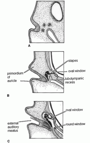
Figure 140.2 Development of the middle ear and ear canal. A: Week 5. B: Week 10. C: Week 27.


Middle Ear Cavity

The cavity and lining of the middle ear and eustachian tube develop from the expanding terminal end of the first pharyngeal pouch with a small contribution from the second pharyngeal pouch. This is apparent in the fourth week of gestation as the tubotympanic recess, which is positioned against the ectoderm of the infolding branchial groove. In the fifth and sixth weeks, the mesenchyme between the branchial cleft and the developing inner ear has condensations destined to become the ossicles. The tympanic cavity continues to develop as the continuing expansion of the endodermal pouch surrounds the ossicles and their supporting structures. It remains a slit-like structure until the fifth month but begins to expand such that the ossicles lie within an open tympanic space by the eighth month (7). Continuation of the tympanic cavity from the epitympanum into the antrum begins at 22 weeks and is complete at birth. Formation of the mastoid air cell system begins late in fetal life, the antrum is present at birth, and continues throughout childhood. The pattern and extent of pneumatization are highly variable. Pneumatization of the petrous pyramid, present in 30% of temporal bones, does not begin until the third year of life (8). At birth, the mastoid tip is not developed but expands through the tractional effect of the sternocleidomastoid attachment.


Ossicles

The exact origin of the ossicles has long been debated. It is certain that the main source is the neural crest mesenchyme of the first and second branchial arches—Meckel cartilage (first arch) and Reichert cartilage (second arch). The otic capsule has a role in formation of the stapes footplate (9). It is generally agreed that the head of the malleus and the body and short process of the incus are formed from Meckel cartilage and are initially continuous with the cartilaginous mandible. The mandibular branch of the trigeminal nerve is the nerve of the first arch; thus, it supplies the tensor tympani muscle, also a derivative of the first branchial arch. The long process of the incus, handle of the malleus, stapes superstructure, and tympanic surface of the stapes footplate are derived from the Reichert cartilage. The facial nerve is the nerve of the second arch; this supplies the stapedius muscle. The vestibular surface of the footplate is a derivative of the mesoderm of the otic capsule, as is the anular ligament (3) (Fig. 140.3).

The malleus and incus are first formed as cartilaginous models from the sixth week of gestation. They begin to ossify in week 16, and ossification is almost complete by week 30. The stapes appears slightly before the malleus and incus. It is initially ring shaped and penetrated by the stapedial artery, the artery of the second arch, which regresses. By 10 weeks, the stapes has already started to assume the familiar stirrup shape. By the time ossification begins from a solitary center at 16 weeks, the structure is a model of the future stapes. It is reduced in bulk throughout fetal life to develop its slender architectural form.


Maldevelopment

The spectrum of abnormal development in congenital atresia of the external auditory canal parallels the fact that the canal is present, albeit short, then is absent before achieving the adult form. In the most severe cases of atresia, a bony mass replaces the tympanic ring and forms the lateral wall of the middle ear cavity, the condyle of the mandible lying more posteriorly. In membranous atresia, which is less common, a fibrous mass replaces the external auditory canal. The mildest form of abnormality is stenosis of the external auditory canal, common in Down syndrome, which can be difficult to diagnose unless complications such as proximal cholesteatoma caused by trapped debris supervene. Congenital atresia is unilateral in 70% of cases (10).

A further consideration with atresia is the presence of coexisting abnormalities of either the pinna or the middle
ear. Associated auricular abnormality exists in 94% of cases of atresia, and the middle ear is frequently deranged (11), in part because all these structures are derived from the first two branchial arches and the intervening branchial cleft. Consequent are further abnormalities of branchial arch derivatives, such as the mandible. Among children with microtia or anotia, 20% to 40% have an identifiable syndromal malformation, such as hemifacial microsomia, Treacher Collins syndrome (mandibulofacial dysostosis), or Goldenhar (oculoauriculovertebral) syndrome (5, 12). These syndromal malformations are discussed in Chapter 86.


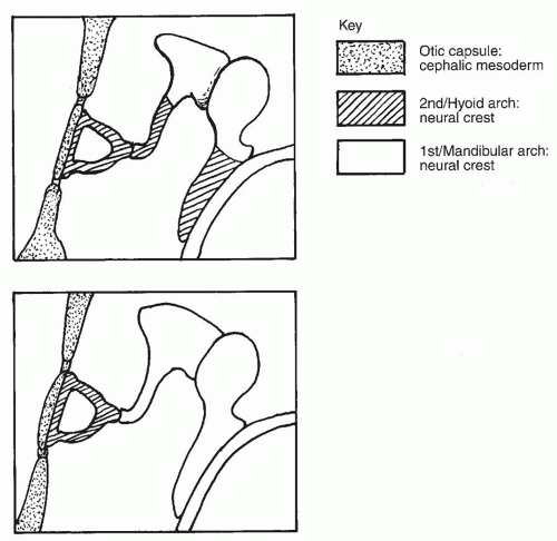
Figure 140.3 Origin of the ossicles—two interpretations.

Ossicular abnormalities also encompass a wide spectrum, from a rudimentary ossicular mass to minor morphologic defects. Middle ear deformities without coexisting outer ear defects are unusual, occurring among fewer than 10% of children with congenital conductive defects (13). This may, however, be an underrepresentation with cases undiagnosed or ascribed to acquired causes. The malleus is always fixed to a bony atretic plate if present, and incudomalleolar fusion or fixation is a common defect. Stapedial abnormalities are less common. In particular, the footplate can have normal mobility, even with a severe coexisting abnormality, because of its separate development from the otic capsule (14).

Persistent stapedial artery is a condition with an interesting embryologic background, although only approximately 50 cases have been reported worldwide. The stapedial artery is the remnant of the second arch artery, which courses from the aortic sac to the dorsal aorta. This artery regresses at approximately 10 weeks’ gestation, and its role is assumed by the precursors of the internal and external carotid arteries. When the artery persists, a vessel arises from the internal carotid artery in the hypotympanum, which courses through the crura of the stapes to the fallopian canal. It enters the fallopian canal and courses forward to the geniculate ganglion and to the dura. If present, a persistent stapedial artery often manifests as a pulsatile mass in the middle ear cavity, pulsatile tinnitus, and rarely, as conductive hearing loss due to stapes ankylosis. The clinical interest is in cases in which middle ear surgery has been undertaken to manage presumptive otosclerosis or for cochlear implantation, as there is an increased risk of bleeding (15). Another condition with an embryologic basis is congenital cholesteatoma. This condition is caused by failure of atrophy of epidermoid formation in the anterior mesotympanum.

May 24, 2016 | Posted by in OTOLARYNGOLOGY | Comments Off on Development of the Ear

Full access? Get Clinical Tree

Get Clinical Tree app for offline access