Introduction
Chronic rhinosinusitis (CRS) unlike acute rhinosinusitis1 is not primarily an infectious disease. It is also associated with other conditions that can contribute to its chronicity. In this chapter we are having a look at these associated conditions such as immunodeficiency, rhinoscleroma, tuberculosis, Wegener’s granulomatosis, non-Hodgkin’s and T-cell lymphomas, gastro-esophageal reflux disease (GERD), diabetes, Long-term steroid usage, congenital abnormalities of ciliary movements or thickened highly viscous nasal mucus.
Chronic rhinosinusitis may or may not be the presenting complaint in many of these cases and a finely tuned threshold of suspicion and awareness of association may be critical to making a diagnosis in these cases. For instance a case of Wegener’s granulomatosis can present with “total unilateral sensorineural hearing loss with associated CRS”. In such a case in our clinical practice the diagnosis was confirmed by C-ANCA and vasculitis diagnosis which led to its proper treatment.
The diagnostic findings and management of CRS in general have been covered in the relevant chapters of this book. In this chapter, the primary emphasis will be on facts regarding the associated conditions. These have been broadly grouped into congenital conditions, tropical diseases, oropharyngeal and gastro-esophageal diseases, granulomatous diseases, viral diseases and other miscellaneous conditions associated with CRS.
Congenital Conditions Associated with Chronic Rhinosinusitis
Primary Ciliary Dyskinesia
This is also known as “immotile cilia syndrome” or “Kartagener’s syndrome”.2 It is a rare, idiopathic, autosomal recessive genetic disorder that causes defective motility of cilia in upper and lower respiratory tract. The impairment or desynchronization of movement of cilia in mucus membrane leads to mucus stagnation and can result in repeated infections of nose, sinuses and lungs. In 38% of these defects, mutations are seen on two genes; DNA11 and DNAH5, both of which code for proteins found in ciliary outer dynein arm.3 This can be associated with situs inversus and the triad of situs inversus, chronic sinusitis and bronchiectasis known as Kartagener’s syndrome.4
The children born with this disease are prone to sinusitis, bronchitis, pneumonia and otitis media. Infertility is also a common finding in these patients. Many patients experience hearing loss, show symptoms of glue ear and may have poor sense of smell. Incidence of this disease is estimated to be 1 in 33000. Clinically the mucus clearance test (sugar or dye) show delayed clearance. Nasal mucosal biopsy and study by electron microscopy confirm the diagnosis.5
There is no cure for this disease. Essentially, optimum medical management and douching of nose with repeated use of appropriate antibiotics slows down the disease. IVF techniques have been used for fertility. Treatment with various chest physiotherapy techniques has been observed to reduce the incidence of lung infections. Lung transplantation has been done in those with severely impaired lung functions.
Young’s Syndrome
It is a disease associated with the occurrence of chronic sinopulmonary infections, persistent azoospermia, normal spermatogenesis and characteristic epididymal findings after exclusion of cystic fibrosis and the immotile cilia syndrome.6 The sperms are normal but unable to flow due to accumulated inspissated secretions in epididymis. The mucus cilia are also normal in respiratory tract.
In 1980, this syndrome was described to be commoner than cystic fibrosis. It was estimated that 1 in 500 males were affected. It was attributed to “mercury poisoning” (pink disease) which was positive in 40% of these cases in USA. The disease is now rarely seen with increasing reduction of mercury usage.
The diagnosis is process of exclusion. It manifests in adolescent age group and patients often present with infertility. The sinus and lung infections are discovered during the investigations. Saccharine test shows delayed mucus transfer, when placed over the inferior turbinate. Emphasis on treatment of nasal condition is on nasal douching and antibiotic use.
Cystic Fibrosis
Cystic fibrosis is a hereditary autosomal recessive disease9 which affects the entire body causing progressive disability and early death.7 It is caused by mutation in the gene for the protein cystic fibrosis transmembrane conductance regulator (CFTR).8 The affected persons are prone to sinus infections, poor growth, diarrhea and infertility.
The condition can be diagnosed by genetic testing, sweat test, blood levels of immunoreactive trypsinogen and increased nasal transepithelial potential difference that leads to decreased chloride secretion and increased sodium and water resorption.10 The hallmark of disease is salty skin.11 Patients have good appetite but poor growth and poor weight gain, excess mucus production, frequent chest infections and coughing, and shortness of breath. The causes of growth failure are multifactorial and include chronic lung infection, poor absorption of nutrients through the gastro-intestinal tract and increased metabolic demand due to chronic illness.
The lung and sinus disease results from clogging of the upper airways due to mucus build-up, poor mucociliary clearance and infections. In the early stages, incessant coughing, copious phlegm production and decreased ability to physical exercise are common. In later stages, pulmonary hypertension, heart failure, hypoxia and respiratory failure requiring ventilator support may occur.
Staphylococcus aureus, Haemophilus influenza and Pseudomonas aeruginosa are the three most common organisms found in lung infections. Opportunist fungal infection by Aspergillus fumigates causes worsening of breathing problem by causing allergic bronchopulmonary aspergillosis.12 Infection with Mycobacterium avium complex (MAC) can cause further lung damage. The paranasal sinus infection may also be seen.13
Churg-Strauss Syndrome
It is also known as “allergic granulomatous angiitis”. This syndrome should be suspected in the presence of any 4 of the 6 features of asthma, migratory pulmonary infiltrates, eosinophilia >10%, para sinonasal abnormalities,14 polyneuropathy and tissue eosinophilia. It is a rare granulomatous vasculitis involving small to medium-sized vessels of unknown cause. The factors implicated are vaccinations, desensitization and various medications like leukotriene receptor antagonist. Approximately 50% have positive P-ANCA on immunoflouroscence with myeloperoxidase (MPO-ANCA) specificity on enzyme immunoassay.15 Other investigations needed are peripheral eosinophil count, ESR, renal function test, chest radiograph, CT scan and ECG.
The most common nasal presentation seen is nasal polyposis (70%), recurrent sinusitis and adult onset asthma. This may later spread in second stage to peripheral blood and tissue eosinophilia, primarily in lungs. In late (third) stage, widespread systemic vasculitis occurs involving peripheral nervous system, skin, heart, gastrointestinal system and kidneys. One of the principal causes of mortality and morbidity in this disease is myocardial disease secondary to coronary arteritis.16
Table 1 Specific Tests and Additional Treatment of Congenital Conditions Associated with Chronic Rhinosinusitis | ||
| Diseases | Specific tests | Additional treatment |
| Primary ciliary dyskinesia | ||
| Young’s syndrome | ||
| Cystic fibrosis | ||
| Churg-Strauss syndrome | ||
Tropical Diseases Associated with Chronic Rhinosinusitis
Rhinoscleroma
It is a rare, chronic, progressive granulomatous disease of upper respiratory tract. The causative agent is Kleibsella rhinoscleromatis.17 Along with other areas, it is endemic in Indian subcontinent. Most frequently it affects adolescents and young adults.18 The organisms multiply intracellularly till mikulicz cell rupture, eventually leading to granuloma formation.19
It can affect any part of upper respiratory tract but manifests primarily in nose. Main symptoms of advanced disease are nasal obstruction (94%), nasal deformity (32%), epistaxis (11%) and nasal crusting (94%).20,21
The diagnosis is based on clinical symptoms, biopsy, culture and CT scan. Treatment includes conservative surgical debridement and long-term antibiotic coverage is the mainstay of therapy. Tetracycline has been shown to be effective unless contraindicated. Flouroquinolones may be used as the alternative medicine. But relapses can occur even with aggressive therapy.
Rhinosporodiosis
The disease is caused by Rhinosporidium seebri.17 It was previously thought to be a rare condition but now it has been reported from several countries of the world.18,22 The modes of infection and transmission of the disease are still not clear. In India it is thought to spread by taking head baths in common ponds with infected water which gets into the nose and causes the infection.23
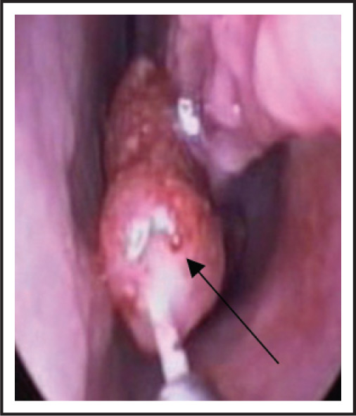
The commonest site of manifestation of rhinosporodiosis in man is the nose (78%). Patients typically present with symptoms of progressive nasal obstruction, chronic epistaxis and watery rhinorrhea which becomes purulent during infection.24 Clinical examination reveals a polypoidal mass, the surface of which contains pin-sized yellow spots giving the mass the classic “strawberry” appearance (Figure 1). The other common sites are pharynx, faucial pillars, soft palate, uvula, larynx and lacrimal sac. It has been observed that rhinosporodiosis restricts itself to mucus membrane and does not cross the muco-cutaneous border.
The treatment is surgical excision with cauterization of the base. Medical treatment has limited efficacy but is done to control the nasal manifestations. This includes saline irrigation, nasal saline and steroid sprays and decongestants. Antifungals and dapsone have also been recommended.
Leprosy
The disease is also known by the name of Hansen’s disease.25 It is caused by the bacteria “Mycobacterium leprae” and “Mycobacterium lepromatosis”. It is primarily a granulomatous disease of the peripheral nerves and mucosa of the upper respiratory tract, though skin lesions are the primary external sign.
Leprosy is not highly infectious and is transmitted via droplet from nose and mouth during contact with untreated cases.26 Those living in endemic areas with inadequate bedding, contaminated water, insufficient diet or other diseases that compromise immune function are at highest risk.
The early signs and symptoms of leprosy are very subtle and occur slowly over years. Numbness, loss of sensations of temperature, touch, pain and deep pressure, painless ulcers, skin lesions, dryness of eyes, loss of digits and facial disfigurement also occurs in the late stages. Nasal symptoms include nasal mucosal lesions, nasal congestion and epistaxis leading to CRS, mainly seen with lepromatous leprosy.
Leprosy can be diagnosed in majority of cases by clinical findings. Smears and biopsy can be done for confirmation. Other tests that are not routinely done are lepromin test, urinary phenolic glycolipid-1 test, PCR, lymphocyte migration inhibition test (LMIT) and antibodies against M. leprae 35-kDa protein (ELISA or the Dipstick method).27
The current treatment recommendation is the use of rifampicin and dapsone for 6 months in paucibacillary leprosy (1–5 skin lesions) and addition of clofazimine for 12 months in multibacillary cases (>5 skin lesions). Saline irrigation and nasal saline and steroid sprays can be used to relieve the nasal symptoms. A single dose of rifampicin and BCG vaccination reduces the rate at which the contacts acquire leprosy.
Tuberculosis
Tuberculosis is one of the oldest diseases known to affect humans. It is caused by bacteria Mycobacterium tuberculosis complex.27 The disease usually affects the lungs although in up to one-third of cases, other organs are also involved.
More than 3.8 million new cases of TB (pulmonary and extra-pulmonary)—90% of them from developing countries—were reported to WHO in 2001. Recent trend in developing countries like India show stable situation with almost no decline.
The disease is most commonly transmitted from a patient of infectious tuberculosis to others by droplet nuclei aerosolized by coughing, sneezing or speaking. Other routes of infection are through skin and placenta.
Tuberculosis of upper airways is nearly always a complication of advanced cavitary pulmonary tuberculosis. It may involve the larynx, pharynx and epiglottis leading to hoarseness and dysphagia. Nasal tuberculosis could be primary or secondary with symptoms and signs of nasal congestion, stuffiness, nasal crusting, epistaxis and ulcerations in the mucosa ending up in CRS.
The diagnosis can be made on the clinical grounds and confirmation is done by the tests like tuberculin skin test, T spot TB blood test, radiology, smear microscopy, culture and nucleic acid amplification test.28
The standard short course treatment for TB is isoniazid, rifampicin, pyrazinamide and ethambutol for 2 months, then isoniazid and rifampicin alone for further 4 months. For latent TB, the standard treatment is 6–9 months of isoniazid alone. Nasal symptoms are managed with saline irrigation, decongestant nasal drops and saline and steroid nasal sprays.
Table 2 Specific Tests and Additional Treatment of Tropical Diseases Associated with Chronic Rhinosinusitis | ||
| Diseases | Specific tests | Additional treatment |
| Rhinoscleroma | ||
| Rhinosporiodosis | ||
| Leprosy | ||
| Tuberculosis | ||
Oropharyngeal and Gastro-Esophageal Diseases Associated with Chronic Rhinosinusitis
Gastro-esophageal Reflux Disease
The disease is defined as chronic symptoms or mucosal damage produced by the abnormal reflux in the esophagus, which can be due to incompetence or transient relaxation of lower esophageal sphincter.
The most common symptoms are heartburn, regurgitation and dysphagia. Other atypical symptoms include chronic cough, laryngitis, asthma, erosion of dental enamel, recurrent ear infections and sinusitis.
A detailed historical knowledge is vital for an accurate diagnosis. Useful investigations may include ambulatory esophageal pH monitoring, barium swallow X-rays, esophageal manometry and esophagogastroduodenoscopy.29
Three classes of treatment exist. These include lifestyle modifications (dietary changes, avoidance of smoking, weight reduction), medications (proton pump inhibitors, H2 receptor blocker, antacids) and surgery (Nissen fundoplication). Nasal symptoms are managed by saline irrigation, antibiotics and nasal saline and steroid sprays.
Samter’s Triad
Samter’s triad is a medical condition consisting of asthma, aspirin sensitivity and nasal polyposis. It occurs in middle age and may not be associated with any allergies.
It goes by several other names like acetylsalicylic acid triad, Widal’s triad, Franci’s triad, aspirin triad and non-allergic rhinitis. When present with CRS, it is called aspirin exacerbated respiratory disease (AERD).
Stay updated, free articles. Join our Telegram channel

Full access? Get Clinical Tree


