Oral manifestations of systemic diseases are potential indicators of an array of conditions. Truly the oral cavity is a mirror that reflects and unravels many of the human body’s internal secrets. Some of these manifestations are disease specific and help raise a high degree of suspicion for the alert clinician. Because oral manifestations may accompany many systemic diseases, it is essential that these are appropriately recognized to provide correct diagnosis and referral for treatment and patient care. Multiple entities involving the various areas of the oral cavity like the soft palate, hard palate, tongue, gingiva, oral mucosa, the dentition, periodontium, and the salivary gland tissue have been enlisted. Although this article is not all-inclusive, the authors highlight lesions or conditions that are directly related to or are caused by some of the more common systemic diseases, and hope to provide ample insight for physicians, dentists, and clinicians in otolaryngologic practice.
Hematologic diseases
Iron-Deficiency Anemia
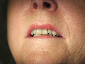
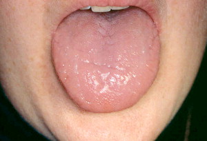
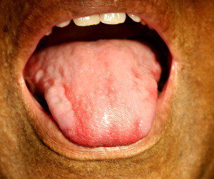
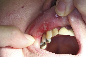
Iron-deficiency anemia is one of the most common causes of anemia worldwide. This type of anemia has a direct relationship between the lack of availability of iron in the body and the demand of red blood cells (RBCs). Oral manifestations may include angular cheilitis, atrophic glossitis, mucosal pallor, and generalized atrophy of the oral mucosa. The glossitis may include flattening of the papillae on the dorsum of the tongue ( Fig. 1 ), and is often accompanied with a tenderness or a burning sensation. This process may result in a smooth, erythematous tongue ( Fig. 2 ) mimicking benign migratory glossitis, also known as “geographic tongue” or erythema migrans; the condition may affect 2% of the population. Geographic tongue may lead to the presence of lesions on the tongue that are red and white, nonindurated, atrophic, but with a slightly elevated, white rim, and the site keeps changing over a period of time. Atrophic glossitis as noted with iron-deficiency anemia, however, does not shift position over time. Angular cheilitis involves the commissures of the lips and causes some degree of cracking. and is generally caused by Candida albicans infection.
Plummer-Vinson syndrome or Paterson-Kelly syndrome is another type of iron-deficiency anemia that presents with glossitis, and is generally seen in women of Scandinavian and Northern European origin. Oral complications include dysphagia, pain on swallowing, presence of abnormal bands of tissue in the esophagus called “esophageal webs,” and spoon-shaped configuration of nails called “koilonychias.” The significance of the condition is that it is premalignant and is related to a high frequency of oral and esophageal squamous cell carcinomas that develop in these patients. It is notable that if the anemia is recalcitrant, host resistance to infection may be significantly lowered.
Pernicious Anemia
Pernicious anemia is an uncommon condition, and recent evidence indicates a slight preponderance in the African American and Hispanic populations in the Unites States. Pernicious anemia is a type of megaloblastic anemia, related to the lack of intrinsic factors in the stomach preventing the absorption of cobalamin (vitamin B12). Symptoms such as fatigue, weakness, shortness of breath, paresthesia, and tingling or numbness of extremities are not uncommon. Moreover, some patients present with memory loss, irritability, and depression.
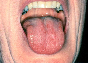
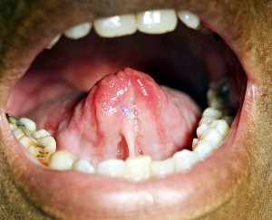
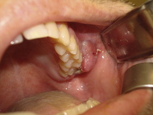
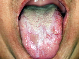
The oral manifestations include burning of the tongue, lips, and buccal mucosa as well as other mucosal sites. Focal or diffuse erythema of the tongue and mucosal atrophy is noted, depending on the duration and severity of the condition. An interesting term, “magenta tongue” ( Fig. 3 ), is often used to describe the presentation. An easy differentiating feature from premalignant conditions that may present in a similar fashion is that the tongue lesions of pernicious anemia are mostly noted on the dorsum ( Fig. 4 ), which is considered a low-risk area for development of squamous cell carcinoma.
Leukemia
Leukemia has a variable presentation and may result in infiltration of the malignant cells in vital organs causing splenomegaly, hepatomegaly, and lymphadenopathy. Most clinical signs and symptoms are related to the reduced numbers of the white blood cells (WBCs) and RBCs. This crisis in turn reduces the oxygen-carrying capacity of the blood resulting in fatigue, easy tiring, and dyspnea. Oral complications of leukemia include petechial hemorrhages of posterior hard palate and soft palate, and spontaneous gingival bleeding. The gingival bleeding is precipitated when the platelet count falls between 10,000 and 20,000/mm 3 . Also noted are mucosal ulcers. Rarely “numb chin syndrome” is appreciated, which is associated with the infiltration of the mental nerve by the malignant cells. Areas of tissue destruction, necrosis, ulceration of the palate, and zygomycosis of the nasal cavity and the paranasal sinuses may be a significant finding. Diffuse oral candidiasis is often a complication of leukemia. Chemotherapeutics during the course of leukemia is one of the triggering factors in reactivation of herpes simplex virus (HSV), leading to the small vesicular type of oral mucositis. Viral herpetic gingivostomatitis may present on both the attached and mobile oral mucosa in contrast to only keratinized mucosa as observed in immunocompetent hosts. The leukemic cells may also produce a diffuse, boggy, nontender swelling with or without ulceration, which appears as a gingival enlargement or even a tumor-like growth containing a collection of leukemic cells called granulocytic sarcoma ( Fig. 5 ) or extramedullary myeloid tumor.
Langerhans Cell Histiocytosis (Histiocytosis X)
Langerhans cell histiocytosis (LCH) is characterized by abnormal monoclonal proliferation of antigen-presenting Langerhans cells. LCH may be focal or disseminated with extensive systemic involvement. LCH represents a spectrum of clinical disorders that are highly aggressive, destructive, and frequently fatal. The common infantile form previously referred to as Letterer-Siwe disease is characterized by involvement of the viscera, and potentially causes death. Skin lesions are common and include papules, plaques, vesicles, and hemorrhagic nodules. The presence of alveolar bone loss in young children with premature primary teeth loss should raise the suspicion of the possibility of LCH. Hand-Schüller-Christian disease is a more localized childhood disease with a triad of diabetes insipidus, lytic bone lesions, and proptosis. LCH can also occur in adolescents and adults. Of the bones of the jaw, the mandible, is the most frequently involved site.
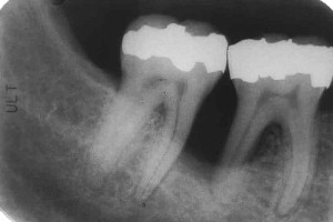
Oral manifestations of LCH include irregular ulcerations of the hard palate, which may be the primary manifestation of the disease. Gingival inflammation and ulcerated nodules, difficulty in chewing, and foul-smelling breath also occur. Large ulcerations with exposed bone, ecchymoses, gingivitis, and periodontitis followed by tooth loss are the hallmark of this condition. Radiographically, the teeth often appear to be “floating in air” and in some cases may present with a large radiolucent region ( Fig. 6 ) exhibiting significant bone loss. These lesions may result in fractures and significant displacement of teeth. Oral swellings or ulcerations resulting from mandibular or maxillary bone involvement may be common, and ulcerations may develop on the gingiva, palate, and floor of the mouth. Sometimes a necrotizing gingivitis-like condition is noted with or without underlying bone destruction.
Multiple Myeloma
Multiple myeloma usually involves the oral cavity in the later stages of the disease. The usual site of involvement is the jaws, mostly the mandible. These lesions may cause significant facial asymmetry and swelling of the jaws ( Fig. 7 ). This condition is associated with bone pain, numbness, mobility of teeth, and at times pathologic fractures. If the disease is limited to only one site and exhibits a monoclonal, neoplastic proliferation of plasma cells in the bone it is termed plasmacytoma, being called extramedullary plasmacytoma when in the soft tissue. Radiographically multiple well-defined punched out lesions or even ragged radiolucencies of the skull and jaw are characteristic findings. An interesting finding is amyloid deposits in the tongue leading to macroglossia, which in some cases prompts a biopsy ultimately leading to the final diagnosis. The periorbital skin may in some cases also exhibit amyloid deposits appearing as waxy, firm, plaque-like lesions.
Autoimmune conditions
Sjögren Syndrome
Sjögren syndrome (SS) is an autoimmune disease, affecting mostly women aged 50 years or older. There is a definite female sex predilection, with a 9:1 ratio as compared with males. SS is characterized by Sicca syndrome or the “primary Sjögren,” comprising xerophthalmia and xerostomia. These SS patients frequently present with a parotid gland enlargement. The secondary form of SS is often associated with another concurrent autoimmune disease, mostly rheumatoid arthritis (RA) or systemic lupus erythematosus (SLE).
The oral manifestations in SS are generally related to the low salivary volume and comprise dysphagia, dysgeusia, difficulty in eating and speaking, as well as an increased rate of dental caries and susceptibility to infection. The saliva is characteristically thick, ropey, and mucinous, or in some severe cases totally absent. The mucosal changes related to xerostomia include dry, red, and wrinkled mucosa. The tongue may appear “bald” or even “cobblestone-like” due to atrophy of the papilla. Cracks, fissures of the tongue, redness, and cheilitis are additional findings. Fungal infestation with Candida albicans is common in persons with SS. These patients also present with a relatively high incidence of dental caries because the amount of saliva is insufficient to dilute dietary sugar. This increase in dental caries is especially noted around the cervical region of the teeth. Inflammation of the parotid gland is a likely complication and is usually accompanied with fever and purulent discharge. Early recognition of SS is important, as this can lead to quick referral of the patient for early management of the dental caries that can progress fairly rapidly. These SS patients are also at greater risk for the development of lymphoma, up to 40 times higher than the normal population.
Kawasaki Disease
Kawasaki disease, also known as mucocutaneous lymph node syndrome, is a systemic vasculitis that affects medium- and large-sized arteries and lymph nodes, and is now considered to be the primary cause of childhood heart disease in the United States. Though rare, if present it is usually noted in children younger than 5 years. An episode of acute edema, erythema of the hands and feet, pyrexia, oral erythema, and multiple rashes are almost always observed. A diagnostic criterion requires that the body temperature must exceed 38.5°C for minimum of 5 days. At least four of these five criteria have to be met to diagnose Kawasaki disease: (1) edema of extremity, erythema; (2) polymorphous exanthem; (3) bilateral conjunctival injection; (4) erythema and strawberry tongue; and (5) acute cervical lymphadenopathy. The most striking oral finding includes swelling of papillae on the surface of the tongue (strawberry tongue) and intense erythema of the mucosal surfaces. The lips may be cracked, red, swollen, and often hemorrhagic and crusted.
Scleroderma (Progressive Systemic Sclerosis; Hide-Bound Disease)
Scleroderma is a rare immunologically mediated condition characterized by deposition of extraordinary amounts of dense collagen in the tissues of the body. Skin is commonly involved, but almost all body organs eventually are affected. There is a 5:1 female predilection.
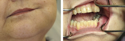
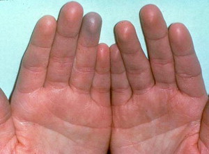
When exposed to cold temperatures these patients show what is termed Raynaud phenomenon in their terminal phalanges. The blood supply to the fingers or toes, and in some cases the nose or earlobes, is reduced, the skin turns pale or white, and numb, and with further depletion of oxygen supply the skin turns blue and cyanotic ( Fig. 8 ). The face appears smooth, taut, stretched, and mask-like, due to subcutaneous collagen deposition ( Fig. 9 ). The “ala” of the nose becomes atrophic and exhibits a pinched appearance called “mouse facies.” Serious sequelae of systemic sclerosis are fibrosis of lungs, heart, kidneys, and gastrointestinal tract, leading to organ failure mostly within 3 years of initial symptoms. Gastrointestinal symptoms such as dysphagia and heartburn are not uncommon.

The oral manifestations are variable. The lips appear pursed due to constriction of the mouth, thus making it difficult to open the mouth and resulting in what is termed microstomia, causing limited opening of mouth in about 70% of these patients. A “purse-string” appearance caused by creases and folds of skin radiating from the mouth may be noted. Xerostomia is a consistent finding, and some of these patients have concurrent secondary SS. The tongue can lose mobility and generally has a smooth appearance. The collagen deposits invariably result in smoothening of the palatal rugae. Diffuse widening of the periodontal ligament space is noted in the entire dentition. The mandible exhibits resorption of posterior part of the ramus, the coronoid process, the chin, and condyles on panoramic radiographs.
Systemic Lupus Erythematosus
SLE is the most common of the so-called vascular collagen disorders in the United States, and more than 1.5 million people remain affected. SLE is an immunologically mediated condition and has multiple clinical presentations. In SLE, apart from the renal and cardiac involvement, oral lesions are noted in 5% to 25% of the patients. The lesions affect the palate, buccal mucosa, and the gingivae; these may present as lichenoid areas that may be nonspecific in appearance, or even sometimes appear granulomatous. “Lupus cheilitis” involving the lower lip vermilion may be noted. Oral ulceration, pain, erythema, and hyperkeratosis as well as complaints of stomatodynia, dysgeusia, xerostomia, candidiasis, and periodontal disease are noted with some frequency in patients with SLE.
Chronic cutaneous lupus erythematosus (CCLE) is limited to the skin and is also known as discoid lupus erythematosus (DLE). In this category the oral lesions are clinically identical to erosive lichen planus ( Fig. 10 ), but these oral lesions are notably absent in the absence of skin lesions. Oral lesions are characteristically ulcerated, atrophic, and erythematous, exhibiting a central zone surrounded by white, fine, radiating striae that are often multiple ( Fig. 11 ). Sometimes these may show a central stippled white dotted area. These oral lesions may be painful when exposed to acidic or salty foods in particular. Oral lesions of the lichen planus are similar to those of DLE both clinically and histologically.
Due to the similar clinical presentation, the lesions of SLE and CCLE must be diagnosed by applying strict histologic criteria, so as to distinguish one from the other. The ulcerations are generally painless and may involve the palate. Salivary gland hypofunction may be concurrently present in patients with SLE, leading to secondary SS and severe xerostomia.
Rheumatoid Arthritis
RA is a chronic, autoimmune disorder causing nonsuppurative inflammatory destruction of the joints. About 3% of the United States population is afflicted by the condition. RA mostly affects women with a 3:1 sex predilection. Some patients have a limited form of the disease with almost no debilitation, pain, or restricted movement. In others there is rapid progress to severely debilitating polyarthralgia.
In the head and neck region the temporomandibular joint (TMJ) is involved to some degree and has been noted in 40% of reported RA patients. This involvement is usually bilateral and is a late finding in the course of the disease. It is usually evident as erosions in the condyle, with subsequent reduced range of motion of the mandible and pain with movement. The condition usually is not as severe as in the other joints that are involved but symptoms may include stiffness, crepitation, pain, or ache. The pain may be a result of pressure and clenching of teeth on one side, thereby inciting pain on the contralateral side. Gross destruction of the mandibular condyle may lead to mandibular micrognathia that presents with a receding chin and distinct malocclusion. Radiographically the condylar head may appear flattened with irregular surface features, and the temporal fossa surface may be eroded and exhibit some remodeling. Many of these patients also have secondary SS.
Amyloidosis
Amyloidosis represents a heterogeneous group of conditions characterized by the deposition of an extracellular proteinaceous material called amyloid. This deposition may be associated with diseases such as multiple myeloma, RA, or chronic infections including tuberculosis. Amyloidosis may be of two types. Organ-limited amyloidosis has rarely been reported in the oral soft tissues. The second type is systemic amyloidosis, which may be in several forms such as primary, myeloma-associated, secondary, hemodialysis-associated, and hereditofamilial.
The primary and myeloma-associated forms of amyloidosis usually affect older adults (average age 65 years). The initial signs and symptoms may be nonspecific. Eventually mucocutaneous lesions and macroglossia develop as a result of the deposition of the amyloid protein. Fatigue, weight loss paresthesia, hoarseness of voice, edema, and so forth may be the first indications of the disease. The skin lesions appear as smooth-surfaced, firm, waxy papules and plaques. Macroglossia has been reported in 12% to 40% of these patients and presents as diffuse or nodular enlargement of the tongue. Sometimes oral amyloid nodules show ulceration and submucosal hemorrhage overlying the lesions. Biopsy of rectal mucosa gingiva and labial salivary gland can be used to confirm the diagnosis.
Autoimmune conditions
Sjögren Syndrome
Sjögren syndrome (SS) is an autoimmune disease, affecting mostly women aged 50 years or older. There is a definite female sex predilection, with a 9:1 ratio as compared with males. SS is characterized by Sicca syndrome or the “primary Sjögren,” comprising xerophthalmia and xerostomia. These SS patients frequently present with a parotid gland enlargement. The secondary form of SS is often associated with another concurrent autoimmune disease, mostly rheumatoid arthritis (RA) or systemic lupus erythematosus (SLE).
The oral manifestations in SS are generally related to the low salivary volume and comprise dysphagia, dysgeusia, difficulty in eating and speaking, as well as an increased rate of dental caries and susceptibility to infection. The saliva is characteristically thick, ropey, and mucinous, or in some severe cases totally absent. The mucosal changes related to xerostomia include dry, red, and wrinkled mucosa. The tongue may appear “bald” or even “cobblestone-like” due to atrophy of the papilla. Cracks, fissures of the tongue, redness, and cheilitis are additional findings. Fungal infestation with Candida albicans is common in persons with SS. These patients also present with a relatively high incidence of dental caries because the amount of saliva is insufficient to dilute dietary sugar. This increase in dental caries is especially noted around the cervical region of the teeth. Inflammation of the parotid gland is a likely complication and is usually accompanied with fever and purulent discharge. Early recognition of SS is important, as this can lead to quick referral of the patient for early management of the dental caries that can progress fairly rapidly. These SS patients are also at greater risk for the development of lymphoma, up to 40 times higher than the normal population.
Kawasaki Disease
Kawasaki disease, also known as mucocutaneous lymph node syndrome, is a systemic vasculitis that affects medium- and large-sized arteries and lymph nodes, and is now considered to be the primary cause of childhood heart disease in the United States. Though rare, if present it is usually noted in children younger than 5 years. An episode of acute edema, erythema of the hands and feet, pyrexia, oral erythema, and multiple rashes are almost always observed. A diagnostic criterion requires that the body temperature must exceed 38.5°C for minimum of 5 days. At least four of these five criteria have to be met to diagnose Kawasaki disease: (1) edema of extremity, erythema; (2) polymorphous exanthem; (3) bilateral conjunctival injection; (4) erythema and strawberry tongue; and (5) acute cervical lymphadenopathy. The most striking oral finding includes swelling of papillae on the surface of the tongue (strawberry tongue) and intense erythema of the mucosal surfaces. The lips may be cracked, red, swollen, and often hemorrhagic and crusted.
Scleroderma (Progressive Systemic Sclerosis; Hide-Bound Disease)
Scleroderma is a rare immunologically mediated condition characterized by deposition of extraordinary amounts of dense collagen in the tissues of the body. Skin is commonly involved, but almost all body organs eventually are affected. There is a 5:1 female predilection.
When exposed to cold temperatures these patients show what is termed Raynaud phenomenon in their terminal phalanges. The blood supply to the fingers or toes, and in some cases the nose or earlobes, is reduced, the skin turns pale or white, and numb, and with further depletion of oxygen supply the skin turns blue and cyanotic ( Fig. 8 ). The face appears smooth, taut, stretched, and mask-like, due to subcutaneous collagen deposition ( Fig. 9 ). The “ala” of the nose becomes atrophic and exhibits a pinched appearance called “mouse facies.” Serious sequelae of systemic sclerosis are fibrosis of lungs, heart, kidneys, and gastrointestinal tract, leading to organ failure mostly within 3 years of initial symptoms. Gastrointestinal symptoms such as dysphagia and heartburn are not uncommon.