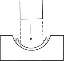
16 See Chapters 3 and 9. Calculate intraocular lens (IOL) power using Sanders-Retzlaff-Kraff (SRK II) formula: Power of IOL = A − 2.5(AL) − 0.9(K), where: 1. Cyclopentolate 1%, phenylephrine 2.5%, and tropicamide 1% every 15 minutes beginning 1 hour before surgery. 2. Optional: Topical nonsteroidal anti-inflammatory agent (e.g., flurbiprofen 0.3% [Ocufen, Allergan, Inc., Irvine, CA, US]) every 30 minutes beginning 2 hours before surgery to minimize intraoperative miosis. 3. Optional: Preoperative antibiotic drops (e.g., moxifloxacin 0.5% [Vigamox, Alcon Laboratories, Inc., Fort Worth, TX, US], gatifloxacin 0.3% [Zymar, Allergan, Inc.]) every 15 minutes for a total of 3 drops may be used as prophylaxis. 1. Anesthesia: Retrobulbar or peribulbar injection plus lid block. May use general anesthesia for younger or un-cooperative patients, hearing or mentally impaired patients, those with language obstacles, or patients with ruptured globes. 2. Decompress eye to avoid positive vitreous pressure. a. Mannitol 20% solution, 250 ml intravenous (slow drip over 1 hour) 1 hour preoperatively. b. Patient to void before entering operating room. c. Secure Honan balloon in position for ~15 minutes (except in cases with globe perforation). 3. Prep and drape. a. Use povidone-iodide 5% on a cotton-tipped applicator to gently clean eyelashes and lid margins. b. Place 1 or 2 drops of povidone-iodide in the conjunctival fornix. Note: There are many types of trephines and corneal punches. The system used is based on surgeon preference. Figure 16.1 4. Trephine donor button from corneoscleral rim (Fig. 16.1). Note: Secure a sterile work area with comfortable surgeon access and adequate lighting, away from the surgical instruments and patient (e.g., work table with stool). a. Hold corneoscleral rim with toothed forceps (e.g., Bishop-Harmon). b. Remove residual fluid from epithelial side of donor to avoid sliding during the trephination (cellulose sponges). c. Place donor tissue epithelial side down (endothelial side up), on Teflon cutting block. d. Trephine appropriately sized button (disposable trephine on universal handle). Note: An 8.0 mm button placed into a 7.5 mm recipient bed is a standard size differential. i. Keep trephine perpendicular to cornea. ii. Punch button in one motion through the entire donor thickness to avoid beveling the edge (listen for “crunch” sound).
Combined Penetrating Keratoplasty/Extracapsular Cataract Extraction/Posterior Chamber Intraocular Lens
Indications
 Patient requiring penetrating keratoplasty for visual rehabilitation of an eye which also has a visually significant cataract
 Patient with symptomatic corneal endothelial dystrophy (e.g., Fuchs) who requires cataract surgery
Preoperative Procedure
 A-constant (A) is determined by the manufacturer for a specific lens. A typical value for a posterior chamber lens is 118.4.
 Axial length (AL) of eye in millimeters.
 Keratometry measurement (K) cannot be directly determined preoperatively.
 The surgeon may use past postoperative keratometry results obtained with a specific technique as an approximate K reading.
 The curvature of the normal fellow cornea may be measured and used in the SRK formula. When using a 0.5 mm oversized graft, however, subtract 1 to 2 diopters from the SRK result as the graft is typically steeper than the original cornea.
Dilate Pupil
Instrumentation
 Honan balloon
 Mannitol 20% solution
 0.12 mm straight Castroviejo forceps
 0.12 mm Colibri forceps
 Bishop-Harmon forceps
 Teflon cutting block
 Marking pen (e.g., methylene blue, gentian violet)
 Cellulose sponges
 Speculum (e.g., Lieberman or Barraquer)
 Kalt or other strong needle holder
 Fine nonlocking needle holder
 Flieringa ring
 Sutures (7–0 Vicryl, 4–0 silk, 10–0 nylon)
 Hemostats
 Radial keratotomy marker
 Disposable trephine (e.g., Storz, Weck)
 Vacuum trephine (e.g., Hessburg-Barron)
 Cautery
 Microsurgical knife (e.g., Superblade, 15 degree, Beaver #75M)
 Viscoelastic substance (e.g., Healon, Amvisc, Viscoat)
 Corneal scissors (right and left)
 Cystotome
 Cyclodialysis spatula
 Lens loop
 Kuglen hook
 IOL forceps
 Muscle hook
 Acetylcholine solution (e.g., Miochol)
 Jeweler’s forceps
 Paton corneal spatula
 Vannas scissors
 McPherson tying forceps
Operative Procedure
 Sinskey hook
Stay updated, free articles. Join our Telegram channel

Full access? Get Clinical Tree


