Purpose
To evaluate the initial changes of normal rabbit corneas after collagen cross-linking (CXL) with a femtosecond laser.
Design
Experimental study in animal eyes.
Methods
The right eyes of 10 male New Zealand white rabbits were treated with CXL with a femtosecond laser. A femtosecond laser was used to create an intrastromal pocket with 80-μm depth and 7-mm diameter. Intrastromal administration of 0.1% riboflavin solution was made into the femtosecond laser–created pocket and the cornea was irradiated with 3 mW/cm 2 ultraviolet A (UVA) light of mean 370 nm wavelength for 10 minutes. The corneal topography and pachymetry were evaluated by the Pentacam. All observations were performed preoperatively and at postoperative day 1, week 1, week 2, and months 1, 3, and 6. Light microscopy was applied to observe changes in the cornea at postoperative month 6.
Results
Cornea healing postoperatively was uneventful in all cases. The central corneal thickness (CCT) in the treated rabbit cornea reached the peak value at postoperative day 1, which descended gradually to the minimum at month 1 and returned to a high level at postoperative months 3 and 6. The CCT data postoperatively was significantly thicker than that preoperatively, except at postoperative month 1 ( P > .05). Steepening of the operative area was found at the corneal front at postoperative day 1, which recovered dramatically after only 1 week. Then the treated area gradually flattened over the course of follow-up. In 8 of 10 rabbits, a demarcation line–like change in the stroma was visible in the Scheimpflug image at slightly increased contrast as early as 1 month after CXL treatment. The micromorphologic examination also confirmed the existence of the demarcation line. Crystalline lens transparency remained unchanged all the time.
Conclusions
CXL with a femtosecond laser appears to be safe. The cornea can realize a faster, uneventful recovery. The stromal demarcation line may be a direct clinical sign to detect an effective corneal cross-linking during the early phase postoperatively.
Riboflavin and ultraviolet light collagen cross-linking of the cornea has been applied clinically as a treatment for keratoconus, post–laser in situ keratomileusis (LASIK) ectasia, and also ectatic disorders such as bullous keratopathy and pellucid marginal degeneration. The standard technique, using ultraviolet A (UVA) at 370 nm wavelength, involves partial or complete central epithelial removal followed by topical administration of riboflavin 0.1% solution to achieve intrastromal penetration.
The femtosecond laser can increase the precision, reproducibility, and safety of the cuts, theoretically. This novel technology has been applied successfully in recent years to enable flap creation in LASIK, the creation of channels for intracorneal rings, and the preparation of donor and host tissue in penetrating and lamellar keratoplasty; it also achieves the arcuate keratotomy for the correction of high astigmatism. An intrastromal pocket could also be created by a femtosecond laser before collagen cross-linking (CXL) to avoid epithelial removal. In an earlier clinical trial, femtosecond laser–assisted CXL has been reported. Here, a similar intrastromal pocket was created by a femtosecond laser before CXL in normal rabbit corneas. The aim of this preliminary study was to investigate the initial corneal morphologic change after this intervention.
Materials and Methods
Animals and Study Design
The right eyes of 10 male New Zealand white rabbits (with body weights of approximately 2.5 kg) were treated. All animals were healthy and free of clinically observable ocular disease. The right eyes were given the collagen cross-linking with a femtosecond laser. Postoperative medications included topical antibiotics (tobramycin 0.3% drops 3 times/day) and steroidal anti-inflammatory drugs (dexamethasone 0.1% drops 3 times/day) for 1 week.
Intrastromal Pocket Creation
The rabbits were premedicated with a subcutaneous injection of diazepam and atropine (1 mg). For general anesthesia, 1.5 mL of ketamine hydrochloride 10% (25 mg/kg body weight) was injected intramuscularly. For additional local anesthesia, oxybuprocaine 0.4% drops were instilled into the right eyes. The femtosecond laser (Visumax, Carl Zeiss Meditec AG, Germany) with the software profile (version 2.50) was used to create a stromal flap with an upper hinge at 80 μm corneal depth, 7 mm diameter centered at the pupil. The spot size was 35 μm and the energy density was 190 nJ at a repetition rate of 200 kHz. Both the spot distance and track distance were 5 μm.
After the flap creation, a mini-spatula was used to enter corneal stroma with a 10-degree side cut and bluntly dissect between the flap and the inferior stroma. Following the pocket creation, a 25-gauge air cannula was introduced through the cut to inject riboflavin. The duration of stromal pocket completion was less than 3 minutes.
Collagen Cross-Linking Procedure
A 0.1-mL dose of 0.1% riboflavin solution was administered with a 25-gauge air cannula into the intrastromal pocket until it was clearly visualized that the entire pocket was bright yellow and stromal infiltration of the riboflavin solution was completed. After 2 minutes, the UVA light exposure was started. An ultraviolet A irradiation source (UV-X; IROC AG, Zürich, Switzerland) of 370-nm wavelength was used to irradiate the corneal surface. The effective fluence at the corneal surface was calibrated at 3 mW/cm 2 for a duration of 10 minutes with the distance between rabbit cornea and UVA light equals to 1 cm.
Preoperative and Postoperative Examinations
Animals were examined before the CXL treatment. Postoperative examinations were performed at postoperative day 1, week 1, week 2, and months 1, 3, and 6. The corneal topography and pachymetry were measured using a rotating Scheimpflug camera (Pentacam; Oculus, Wetzlar, Germany). The anterior segment image was acquired at the same time by the Pentacam. For the corneal pachymetry, the central corneal thickness (CCT) was manually measured at the 90-degree image by the same examiners. Three consecutive measurements were performed, from which the median data were adopted.
Histologic Methods
The micromorphologic examination was performed at postoperative month 6. Corneoscleral tissues were dissected from eye globes, followed by fixation in 4% formaldehyde for approximately 48 to 72 hours and then embedded in paraffin. The sections of 4-μm thickness were stained with hematoxylin-eosin (HE).
Statistical Analysis
Statistical analysis was performed using Microsoft Excel 2007 (Microsoft; Redmond, Washington, USA) and SPSS 13.0 software (SPSS Inc, Chicago, Illinois, USA). Comparisons between preoperative and postoperative data were performed by paired t tests. A P value < .05 was considered statistically significant.
Results
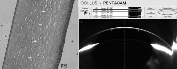
All rabbits could open their eyelids naturally and return to full activities within 1 day postoperatively. No negative biomechanical effect was observed attributable to the femtosecond laser–created pocket. Cornea healing postoperatively was uneventful in all cases. Steepening of the operative area was found at the front at postoperative day 1, which recovered dramatically after only 1 week. Then the treated area went on to flatten gradually, with decrease of the keratometry (K) readings in the follow-up ( Figure 1 ). In 8 of 10 rabbits, a demarcation line–like change in the stroma was visible in the Scheimpflug image at slightly increased contrast as early as 1 month after CXL treatment. The stroma before the demarcation line was higher than the posterior in reflection density and the depth of CXL was thicker in the center than in the periphery ( Figure 2 ). The micromorphologic examination at postoperative month 6 also confirmed the existence of the demarcation line between cross-linked stroma and the untreated stroma ( Figure 2 ). A lacunar hydration pattern can be observed on the anterior side of the demarcation line. Transparency of crystalline lens remained unchanged all the time.
One animal died just before the 6-month examination; the death was not related to the CXL intervention. In the other 9 rabbits, the initial CCT in the treated rabbit cornea was 318.9 ± 9.3 μm. It reached the peak value at postoperative day 1 and descended gradually to the minimum value at month 1. Similar data were acquired at both month 3 (351.1 ± 11.7 um) and month 6 (348.9 ± 7.8 um) postoperatively ( Table 1 ). The CCT data postoperatively was significantly thicker than that preoperatively, except at postoperative month 1 ( P > .05) ( Figure 3 ).
| Animal No. | Before | 1 Day | 1 Week | 2 Weeks | 1 Month | 3 Months | 6 Months |
|---|---|---|---|---|---|---|---|
| 1 | 330 | 360 | 350 | 350 | 310 | 350 | 340 |
| 2 | 330 | 360 | 360 | 340 | 310 | 340 | 340 |
| 3 | 310 | 370 | 350 | 330 | 320 | 360 | 350 |
| 4 | 310 | 360 | 330 | 320 | 300 | 350 | 350 |
| 5 | 320 | 350 | 340 | 330 | 310 | 330 | 340 |
| 6 | 310 | 360 | 330 | 320 | 300 | 350 | 350 |
| 7 | 310 | 370 | 350 | 340 | 320 | 360 | 360 |
| 8 | 330 | 380 | 360 | 340 | 320 | 350 | 350 |
| 9 | 320 | 380 | 370 | 350 | 340 | 370 | 360 |
| Mean ± standard deviation | 318.9 ± 9.3 | 365.6 ± 10.1 | 348.9 ± 13.6 | 335.6 ± 11.3 | 314.4 ± 12.4 | 351.1 ± 11.7 | 348.9 ± 7.8 |
Stay updated, free articles. Join our Telegram channel

Full access? Get Clinical Tree


