Fig. 2.1
Disc photograph of a 16-year-old male patient with crescent-type peripapillary chorioretinal atrophy (PPA) who was followed-up for 6 years. (a) Baseline disc photograph shows a small area of PPA and a large disc. (b) Two years later, an enlarged PPA and disc tilt were observed. Cilioretinal vessels in the 8 o’clock sector (arrowhead) remained at the prior disc margin. (a) Six years later, a larger area of PPA and more prominent disc tilt were observed. Note the progressive nasalization and the nasal rotation of the central retinal vessel trunk [23]
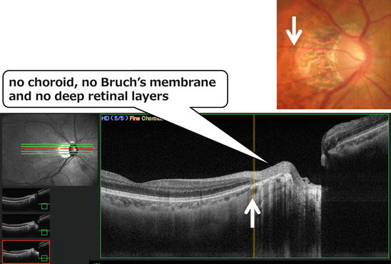
Fig. 2.2
Gamma zone PPA, spectral domain optical coherence tomography images. In the gamma zone, only the retinal nerve fiber layer bundle and the underlying scleral tissues are apparent, with very short terminal fragments of retinal layers and choroidal tissues observed just inside the gamma zone margin (white arrows)
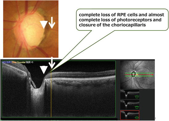
Fig. 2.3
Beta zone PPA, spectral domain optical coherence tomography images. In the beta zone, thin choroidal tissues are visible up to the disc margin (arrowhead). Retinal layers also extend to the disc margin in a tapering configuration across the limits of the PPA (arrows)
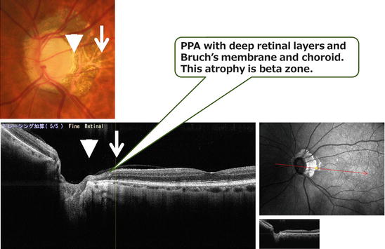
Fig. 2.4
Hard differentiation by the color tone of the PPA area in fundus photographs. Although this disc photograph appears as gamma zone PPA, the OCT image suggests the PPA is beta zone, with deep retinal layers and Bruch’s membrane and choro
2.7 Conclusions
There are many myopic glaucoma cases where both myopic changes and glaucomatous changes are thought to be present. Ophthalmologists should manage myopic glaucoma patients during relatively early stages before the development of impaired visual acuity and central visual field defects. The difference in structural changes in the optic disc, including lamina cribrosa and its surroundings, between myopic and non-myopic glaucomatous eyes may affect the frequency of disc hemorrhage and deterioration rate of the visual field loss. The myopic conus lacks Bruch’s membrane corresponding to the gamma zone, and glaucomatous PPA has Bruch’s membrane corresponding to the beta zone.
References
1.
2.
Nitta K, Saito Y, Sugiyama K (2006) The influence on the static visual field of peripapillary chorioretinal atrophy-relation to axial length. Nihon Ganka Gakkai Zasshi 110(4):257–262PubMed
3.
Ohno-Matsui K, Shimada N, Yasuzumi K et al (2011) Long-term development of significant visual field defects in highly myopic eyes. Am J Ophthalmol 152(2):256–265. e1. doi:10.1016/j.ajo.2011.01.052 CrossRefPubMed
4.
Curtin BJ (1985) Basic science and clinical management. In: Curtin BJ (ed) The myopias. Harper & Row, New York, pp 301–308
5.
Ohno-Matsui K, Akiba M, Moriyama M et al (2011) Imaging the retrobulbar subarachnoid space around the optic nerve by swept-source optical coherence tomography in eyes with pathologic myopia. Invest Ophthalmol Vis Sci 52(13):9644–9650. doi:10.1167/iovs.11-8597 CrossRefPubMed
6.
Ohno-Matsui K, Akiba M, Moriyama M et al (2012) Acquired optic nerve and peripapillary pits in pathologic myopia. Ophthalmology 119(8):1685–1692. doi:10.1016/j.ophtha.2012.01.047 CrossRefPubMed
7.
Akagi T, Hangai M, Kimura Y et al (2013) Peripapillary scleral deformation and retinal nerve fiber damage in high myopia assessed with swept-source optical coherence tomography. Am J Ophthalmol 155(5):927–936. doi:10.1016/j.ajo.2012.12.014 CrossRefPubMed
Stay updated, free articles. Join our Telegram channel

Full access? Get Clinical Tree


