Clinical Evaluation of the Patient with Vertigo


Clinical Evaluation of the Patient with Vertigo


Courtney C. J. Voelker

Joel A. Goebel


INTRODUCTION


Magnitude of the Problem in Population

The diagnosis and management of the patient with dizziness and imbalance can be perplexing and challenging. Dizziness is a common symptom affecting 20% to 30% of the general population in the United States (1, 2). Among patients 75 years of age and older, balance disorders are the most common reason for visiting a physician (3). In the elderly, falls caused by dizziness and poor balance are associated with morbidity and even mortality. Such falls constitute a major health care burden in our aging population (4). Early detection and intervention in these cases can result in significant improvement in the quality of life as well as in a decrease in disability and reduction in health care costs (4). It is, therefore, incumbent on those who diagnose and treat dizziness to recognize the complexity of the problem, develop a systematic and efficient process for gathering accurate historical information, perform a structured neurotologic examination, and provide a rational treatment approach that addresses all major contributing factors.


Multifactorial Nature of Dizziness

The three primary sensory inputs responsible for balance originate from the vestibular, visual, and somatosensory (proprioceptive) pathways. Maintenance of balance requires accurate integration of three processes: (a) appropriate detection of various environmental sensory inputs, (b) accurate central nervous system (CNS) integration of all sensory input and production of an appropriate neural response, and (c) performance of the correct muscle response for maintenance of postural control and gaze stability (Fig. 165.1). The vestibular labyrinth can be divided into the semicircular canals (SCCs) (horizontal, superior, and posterior) and the otolith organs (utricle and saccule). The primary function of the SCCs is to detect high-velocity, angular head movement and to produce a compensatory eye movement via the vestibuloocular reflex (VOR) for stabilization of visual images on the retina and maintenance of acuity during head movement. In contrast, the primary function of the otolith organs is to sense linear acceleration (including gravity) and to produce compensatory postural changes in response to transient linear movements and changes via gravity through the vestibulospinal reflex (VSR) pathways. Taken together, the VOR and VSR serve to stabilize the visual world and upright stance and ambulation, respectively.

The visual tracking and fixation pathways sense movement in the visual surround and allow the individual to respond to either slowly moving (smooth pursuit, optokinetic) or novel (saccadic) objects of interest and augment the VOR for maintenance or acquisition of desired targets. With the vestibular system acting as the “gold standard” internal frame of reference, visual inputs are interpreted in combination with vestibular cues for generation of the desired eye movement for the task at hand.

The somatosensory inputs from the feet, lower limbs, and trunk serve to orient the body to contact surfaces and respond to transient perturbations by generating reflexive corrective limb movements using the ankle joint as the primary fulcrum of movement during upright stance. These cues, along with vestibular inputs, are critical for the maintenance of upright stance and gait control. The absence of accurate vestibular cues or the transmission of incorrect cues invariably leads to poor posture control and ambulation, often resulting in falls.

The second component of balance control is sensory integration, which requires the brain to interpret and weigh all sensory inputs and generate a rapid and accurate response (Fig. 165.1). In some instances, sensory cues are in conflict or ambiguous. Under these circumstances, the brain must decide which cues are appropriate for the given situation and ignore or suppress those that have an incorrect orientation. In susceptible individuals, motion sickness can result.


Figure 165.1 An integrated approach to the diagnosis of dizziness and imbalance. Maintaining balance requires a patient to sense the environment (via sensory afferent input), to integrate the environmental information via central processing, and to calculate the correct motor response (delivered via motor efferent output). Accurately locating the abnormality within this integrated system is crucial when a patient presents with dizziness, imbalance, or vertigo. (From Hughes GB, Pensak ML, eds. Clinical otology, 2nd ed. New York: Thieme Medical Publishers, 1997:44, with permission.)

The final component of posture control is generating an appropriate motor response to sensory inputs. This elaborate task requires an intact peripheral motor system and adequate musculoskeletal capability. In some cases, sensory input and central integration are intact, but inadequate motor capabilities lead to imbalance and falls. It is, therefore, important in each patient with imbalance or dizziness to decide whether or not there is a sensory deficit, a central integration problem, an inadequate musculoskeletal response, or a combination of factors leading to their symptom complex.


Importance of History

A structured and thorough discovery of all pertinent historical events is critical in the evaluation of the dizzy patient (5). In roughly 75% of cases, the history alone yields an accurate differential diagnosis, even before the physical examination or laboratory tests are performed (5). The reasons a precise history is essential are as follow: (a) Most disease processes that cause dizziness have a particular symptom pattern; (b) diseases of the inner ear and eighth cranial nerve cause distinctly different sensations than CNS, musculoskeletal, systemic, or psychogenic processes; (c) most patients are not experiencing an acute sensation of dizziness at the time of examination; and (d) the CNS has a remarkable way of compensating between episodes such that clinical examinations and laboratory findings are only seen during acute episodes or in cases of significant damage. As discussed below, development of a systematic approach to history taking allows the examiner to build a differential diagnosis even prior to performing the physical examination and further testing.


Evaluation of Sensory (Vestibular, Visual, and Somatosensory), Central Integration, and Motor Control Aspects

After obtaining a comprehensive history, the examiner then proceeds to perform a basic otolaryngologic and structured neurotologic examination. Causes of dizziness should not be overlooked at this stage and must be detected on the routine otolaryngologic head and neck examination (e.g., impacted cerumen, middle ear effusion, obstructed nasal airway, and sinus disease). Following this examination, a detailed evaluation of spontaneous and evoked eye movements, VOR and VSR functions, posture, and gait is performed. Special attention is paid to the function of each sensory input, central integration of these inputs, and the generation of an appropriate eye or trunk/limb movement for maintenance of gaze and upright stance.


Judicious Use of Laboratory Testing to Quantify Deficits

In many cases, the etiology of the patient’s dizziness is clear subsequent to taking the history and performing a thorough physical examination. Further audiovestibular, radiologic, and serologic testing may be necessary to support the suspected diagnosis and to quantify the extent of the deficit prior to initiation of medical and/or surgical management. Furthermore, vestibular function tests as a whole exhibit fairly high specificity (80% to 85%), but only modest sensitivity (60% to 65%) (6). Consequently, vestibular function tests are limited in their ability to “rule out” dysfunction for most diseases affecting the labyrinth or eighth cranial nerve. It is, therefore, incumbent on the specialist who manages the dizzy patient to understand the critical role of taking a comprehensive history and performing a structured physical examination prior to ordering any further tests.


HISTORY TAKING


Methods: Questionnaire and Direct Questioning

A variety of methods exists for gathering historical information from the patient prior to the physical examination: (a) structured questionnaire mailed to the patient (see included example), (b) phone interview and verbal


gathering of information, and (c) face-to-face interview at the time of the physical examination. The advantage of a structured questionnaire is its capability to supply comprehensive data regarding the nature of the dizziness/imbalance episodes, accompanying symptoms, additional medical conditions, medications, and lifestyle. However, such questionnaires must be simplified so that it is understandable for the patient and well organized for the practitioner to efficiently interpret the data. Moreover, these questionnaires should be completed prior to the appointment rather than hurriedly filled out in the waiting room. A well constructed and thoughtfully completed questionnaire can be a valuable tool for the examiner to review prior to the patient interview. Alternatively, a medical assistant can conduct a phone interview and record the data. The disadvantages of this approach include that this method can be more time consuming and costly, and the resulting data are less comprehensive than the data from a written questionnaire. Finally, the face-to-face interview by the physician is the most important step in confirming and/or clarifying what the patient has either written in the questionnaire or told the medical assistant by phone. Using the written questionnaire as a guide allows the examiner to ask directed questions during the interview in an efficient fashion to best understand the patient’s symptom complex (Table 165.1).


image


image


image


image


Components of the History


Distinction Between Vertigo, Disequilibrium, and Light-Headedness

The first important piece of information in the history is the nature or quality of the sensation experienced during their episodes. Types of dizziness can be roughly divided into three broad categories: (a) vertigo, (b) disequilibrium, and (c) light-headedness (Table 165.1). Vertigo can be defined as a false sense of motion within the patient’s environment and is usually described as a “spinning, whirling, tumbling, or even rhythmic rocking” feeling (7). In most instances, patients will sense that the environment is in motion around them (objective vertigo) and may describe the illusion that objects within their visual world (e.g., pictures, furniture, etc.) are actually moving in one direction or another before their eyes. In a minority of cases, the patients feel that they are in motion relative to a stationary world (subjective vertigo). In either event, the illusion of motion is distinct. Such strong sensations of environmental and/or self-motion are most often generated by a sudden asymmetry within the peripheral vestibular system (labyrinth or eighth cranial nerve) and are valuable indicators of peripheral inner ear or nerve dysfunction.


TABLE 165.1 THE HISTORY OF PRESENT ILLNESS IN A PATIENT WITH DIZZINESS


Components of the Interview


Key Historical Features


What is the character of the dizzy sensation?


Vertigo, disequilibrium, or light-headedness


What is the dizziness pattern?


Continuous or episodic


What is the time course of dizzy episodes?


Seconds, minutes, hours, days, or longer


Was there an event associated with dizziness onset?


Head/inner ear trauma, barotrauma, upper respiratory infection, ear infection, systemic illness or infection, ototoxic medication


What associated symptoms accompany the dizzy episodes?


Hearing loss, tinnitus (continuous vs. pulsatile), aural fullness, conductive hyperacusis, diplacusis, dysacusis, autophony, oscillopsia, otorrhea, otalgia, headache, facial or limb weakness, dysphagia, dysphasia, visual changes, photophobia, phonophobia, loss of consciousness, seizure


What exacerbates the dizziness?


Rapid head movement, particular head positions, increased pressure (Hennebert sign), sounds (Tullio phenomenon), hyperventilation, strong environmental stimuli (bright lights, odors, etc.), food triggers


What medications (past and present) could be involved?a


Antibiotics, antineoplastic, analgesics, antihypertensives, neuroleptics, antidepressants, sedatives


What is the patient’s past medical history?


Migraine, endocrine (e.g., diabetes), rheumatologic, cancer, cardiovascular, systemic infections


What is the patient’s past surgical history?


Focusing on otologic or brain surgery


What is the family history?


Migraine, endocrine (e.g., diabetes), rheumatologic, cancer, cardiovascular, systemic infections, genetic mutations


a See Table 165.2 for a more detailed discussion.


In contrast with vertigo, patients who have disequilibrium describe difficulty maneuvering within their physical environment often without experiencing an illusion of motion (Table 165.1). Patients with disequilibrium may use synonyms like “imbalanced, clumsy, uncoordinated, or fear of falling,” and their symptoms are usually worse while standing or ambulating. A variety of CNS, peripheral neuropathic, and musculoskeletal disorders can cause disequilibrium, although slowly progressive, bilateral vestibular loss without significant asymmetry may present with this complaint.

The third category—light-headedness—comprises both a wide range of sensations (such as “wooziness, giddiness, feeling faint, or as if one is about to pass out”) and of etiologies (including migraine, vascular, metabolic, druginduced, endocrine, or primary psychogenic causes) (Table 165.1). Light-headedness, though, is less commonly a primary complaint of patients with damage of the peripheral vestibular pathways. Instead, light-headedness is most often comorbid with vertigo and/or disequilibrium.


Time Course

Once the nature and quality of the sensation is elucidated, the examiner then attempts to determine the duration of the symptoms (Table 165.1). The first question is whether or not the sensation is continuous or episodic and, if so, do the episodes last seconds, minutes, hours, days, or even longer. When patients complain of vertigo, this time course distinction is exceedingly valuable since the most common peripheral vestibular disease processes produce stereotypical attacks of consistent duration. For example, patients with benign paroxysmal positional vertigo (BPPV) usually complain of discrete attacks that last less than 1 minute. In contrast, patients with Ménière’s disease usually describe a “spinning feeling” that can last 15 minutes to many hours. Finally, patients who develop vertigo secondary to vestibular neuritis describe attack(s) of continuous spinning lasting up to 24 hours. In patients with transient ischemic attacks involving the vertebrobasilar circulation and brainstem, attacks generally last 15 minutes, whereas brainstem infarcts and cerebellar hemorrhages exhibit acute, severe vertigo lasting hours and impact the inability to stand.

The time course of attacks in patients with disequilibrium is somewhat less stereotypical of their disease process. Frequently, patients report that symptoms persist as long as they are upright. Similarly, patients with light-headedness may have a variable symptom picture, with sensations lasting anywhere from seconds to days. Finally, patients with migraine-associated dizziness (MAD) may complain variably of all three sensations, and the duration may fluctuate from seconds to days or even longer.


Associated Events

It is crucial to elucidate associated events that occurred near the time the dizziness began, such as trauma, recent or chronic infection(s), systemic diseases (e.g., rheumatologic, autoimmune, or metabolic), or medication changes. Posttraumatic vertigo can be the result of (a) direct mechanical trauma to the head or inner ear structures (e.g., penetrating injuries damaging the inner ear, SCC dehiscence) and/or (b) barotrauma (i.e., damage caused by pressure changes). Activities associated with barotrauma may include military service with exposure to blast injuries, scuba
diving, hyperbaric oxygen treatments, straining (e.g., weight lifting or childbirth), or changes in altitude (e.g., airplane flights or driving in the mountains), especially during an upper respiratory infection that causes congestion. Posttraumatic vertigo may be due to perilymphatic fistula(s) (PLF) of the oval and/or round windows (8, 9, 10, 11), delayed endolymphatic hydrops (12), BPPV (13), migraine, or middle ear surgery (14). Furthermore, trauma may cause a dehiscence of the superior SCC if the bone covering the canal was thin prior to the injury (15, 16, 17).

Determining a history of recent infections is important. For example, vestibular neuritis is thought to be caused by viral infection, and an upper respiratory viral prodrome may occur prior to vertigo onset (18). Other infections that can cause vertigo include herpes zoster oticus (Ramsay Hunt syndrome), suppurative otitis media, human immunodeficiency virus, syphilis, Lyme disease, and tuberculosis. Additionally, recent systemic infections or cancers may have necessitated the administration of ototoxic drugs that can cause vertigo.

Finally, it is imperative to determine if the patient also has a systemic condition including a rheumatologic, autoimmune (e.g., Wegener granulomatosis, systemic lupus erythematosus), and/or metabolic (e.g., diabetes) disease that may be associated with vertigo.


Exacerbating Factors

Since the vestibular labyrinth is sensitive to angular motion (turning the head), transient linear motion (sudden translations), and gravity (tipping the head and body), disorders of the inner ear are frequently aggravated by head movements and changes of the head and/or body vis-a-vis gravity (Table 165.1). Patients with vestibular disorders tend to keep their head as still as possible and avoid sudden movements. Vertigo induced by very specific head positions (e.g., rolling over in bed, or tilting the head back to rinse their hair in the shower), which lasts for seconds, suggests BPPV. In cases of bilateral and uncompensated unilateral vestibular dysfunction, patients have difficulty walking in the dark or on uneven surfaces and avoid fast head movements due to bobbing or blurring of their visual field (oscillopsia). Patients with orthostasis become more symptomatic with changes in position against gravity such as arising from a bed or chair and obtain relief by lying flat.

Patients with superior SCC dehiscence may present with sound-induced vertigo (Tullio phenomenon) or pressureinduced vertigo (Hennebert sign) (17, 19). Valsalva maneuvers, coughing, or sneezing can induce vertigo or oscillopsia. Furthermore, hyperventilation (e.g., exercising) may induce vertigo in peripheral (e.g., cerebellopontine angle [CPA] tumors) or central (e.g., multiple sclerosis) lesions.

In cases with MAD, exposure to visual motion, bright lights, loud sounds, or strong odors can exacerbate dizziness. Furthermore, MAD patients can, at times, identify food triggers such as caffeine, alcohol, cheese, or citrus fruits. Changes in dietary salt intake may trigger dizziness in patients with Ménière’s disease.


Medications and Comorbidity

Dizziness is a common side effect of many medications, and, in some instances, the interaction of multiple medicines is the cause of a patient’s symptoms (Table 165.2). Medications causing dizziness, light-headedness, and/or auditory symptoms generally are divided into three categories: those that are (a) ototoxic, (b) affect systemic blood flow, and (c) act on the CNS. Once the drug is stopped or the dose adjusted, symptoms may reverse. However, in some circumstances, the damage may be permanent. Therefore, the examiner must query the patient not only about current medications but also about previous medication usage.

Ototoxic drugs damage the peripheral vestibular system. Ototoxicity usually results in a symmetric loss of vestibular end organ function, which disables the VOR. Thus, patients loose visual stability with head movement, resulting in oscillopsia (i.e., bobbing or jiggling of the visual field). Patients with bilateral vestibular loss present with disequilibrium and have tremendous difficulty or are completely unable to maintain balance and posture in the dark. The most common ototoxic drugs are aminoglycosides and chemotherapy agents (Table 165.2). Aminoglycoside antibiotics (e.g., streptomycin, gentamicin, neomycin, tobramycin, amikacin, kanamycin) constitute a group of natural and semisynthetic compounds used to treat aerobic gramnegative bacilli and mycobacterium. Aminoglycosides inhibit bacterial protein synthesis by binding to the
30S ribosomal subunit and cause mRNA to be misread. While the use of systemic aminoglycosides is declining in developed nations due to their significant toxicities and the availability of better alternatives, aminoglycosides are still widely used in developing countries. This is because aminoglycosides are inexpensive and effective against diseases such as multidrug-resistant tuberculosis (20, 21).


TABLE 165.2 MEDICATIONS THAT MAY CAUSE DIZZINESS


Drug Class


Mechanism


Symptoms


Ototoxic


Aminoglycosides Gentamicin, neomycin, tobramycin, amikacin, kanamycin


Damages vestibular type I sensory cells, outer hair cells in the organ of Corti, and cochlear and vestibular neurons Loop diuretics and vancomycin can potentiate ototoxicity


Oscillopsia from symmetric vestibular hypofunction Vertigo from asymmetric vestibular loss, sensorineural hearing loss, and tinnitus


Antineoplastic agents Cisplatin


Damages vestibular hair cells, outer hair cells in the organ of Corti, stria vascularis, and spiral ligament


Same symptoms as aminoglycoside damage


Analgesic agents NSAIDs (aspirin, naproxen, indomethacin, and ibuprofen)


Reversible outer hair cell function changes and reduced blood flow via vasoconstriction


Reversible sensorineural hearing loss, tinnitus, and rarely vertigo


Acetaminophen/hydrocodone


Permanent hair cell damage


Rapidly progressive, permanent sensorineural hearing loss, tinnitus, and rarely vertigo


Systemic Blood Flow


Antihypertensives Diuretics, β-blockers, vasodilators, CCB α-adrenergic blocker


Systemic vasodilation leading to decreased peripheral vascular resistance


Light-headedness, syncope, visual changes, and fatigue


Central Nervous System


Neuroleptics Antidepressants Tricyclics, MAOIs, SSRIs sedatives


Target central dopamine, serotonin, GABA, and acetylcholine neurotransmitter pathways


Light-headedness, vertigo, and ataxia


CCB, calcium channel blocker; GABA, γ-aminobutyric acid; MAOIs, monoamine oxidase inhibitor; NSAIDs, nonsteroidal anti-inflammatory drugs; SSRI, selective serotonin reuptake inhibitor.


All aminoglycosides cause cochlear and vestibular damage that is usually permanent. However, individual aminoglycosides differ in their ability to produce cochlear versus vestibular toxicity. Gentamicin and streptomycin are primarily vestibulotoxic (22), whereas neomycin, amikacin, and kanamycin are more cochleotoxic. Aminoglycosides destroy vestibular type I sensory hair cells, outer hair cells in the organ of Corti (from cochlear base to apex), and cochlear and vestibular neurons (20, 21). The perilymph and endolymph drug concentration is directly proportional to the plasma concentration, which in turn is directly related to renal clearance (23). Aminoglycosides persist in the inner ear tissue for 6 months or longer after administration (24). The ototoxic damage may be potentiated by concurrent administration of loop diuretics (e.g., ethacrynic acid and furosemide) or vancomycin. Patients with a mutation in the mitochondrial 12S ribosomal subunit (MTRNR1) are particularly susceptible to aminoglycoside ototoxicity. This mutation is associated with spontaneous as well as aminoglycoside-induced hearing loss even following a single dose. Interestingly, the vestibular system does not have an increased susceptibility to aminoglycoside toxicity in patients with this mutation (25). In contrast, missense polymorphisms in three oxidative stress-related genes (NOS3, GSTZ1, and GSTP1) have increased susceptibility to gentamicin-induced vestibular dysfunction (26).

Numerous chemotherapeutic drugs are ototoxic. The alkylating agent cisplatin (cis-diamminedichloroplatinum II) is the most ototoxic antineoplastic drug and is used to treat various malignancies including head and neck cancers. Cisplatin targets the outer hair cells in the organ of Corti, stria vascularis, and the spiral ligament (Table 165.2) (27). Vestibular toxicity seems to occur later than auditory toxicity. Both elderly and pediatric patients are reportedly more sensitive to cisplatin ototoxicity than other age groups (28). Patient symptoms of bilateral vestibular loss will present in the same way as aminoglycoside toxicity.

There is evidence that high-dose analgesic use is ototoxic. Nonsteroidal anti-inflammatory drugs (NSAIDs) such as salicylates (aspirin) (29), naproxen (30), indomethacin, and ibuprofen are among the offending agents (31, 32). High-dose salicylates (several grams per day) cause outer hair cell dysfunction and decreased blood flow to the inner ear via vasoconstriction, possibly mediated by antiprostaglandin activity. The result is reversible hearing loss and tinnitus (29, 33, 34). Other NSAIDs have similar ototoxic effects through reduction of blood flow (29). Acetaminophen/hydrocodone (e.g., Vicodin, Lortab,
and Norco) abuse has been reported to cause permanent, rapidly progressive, sensorineural hearing loss (35, 36, 37, 38). Although less common than auditory symptoms, vestibular symptoms (e.g., vertigo or disequilibrium) caused by high-dose analgesics can occur. When vestibular symptoms do develop, their onset can even precede tinnitus (Table 165.2) (39).

Many types of antihypertensive medications can cause dizziness (Table 165.2). Examples include diuretics, β-blockers, vasodilators, calcium channel blockers, and α-adrenergic blockers. These medications result in dizziness through persistent orthostatic hypotension or, in part, by effecting CNS neurotransmitter pathways. Symptoms can include light-headedness, syncope, visual changes, and fatigue. Often these symptoms improve or resolve completely with dose adjustments.

Drugs acting on the CNS can cause dizziness (Table 165.2). The pharmacologic targets of neuroleptics, antidepressants (e.g., tricyclics, monoamine oxidase inhibitors, and selective serotonin reuptake inhibitors), and sedatives are located in the cortex or the brainstem. Several of these drug classes affect neurotransmitters such as dopamine, serotonin, acetylcholine, and γ-aminobutyric acid (GABA), which can lead to hypotension, light-headedness, vertigo, and ataxia. At therapeutic doses, Dilantin can often lead to ataxia, vertigo, and gaze-evoked nystagmus (GEN). The first step in diagnosis is obtaining a detailed medical history, including a thorough medication history, and determining the character of the dizziness. Medication side effects may be the sole offender or may be exacerbating an underlying vestibular pathology.


TABLE 165.3 THE NEUROTOLOGIC EXAMINATION OF THE DIZZY PATIENT


Examination


Test Performance


Outcome


Interpretation of Clinical Finding


Spontaneous nystagmus


Static visual fixation


Nystagmus waveform, direction, effect of fixation


Peripheral: Horizontal-rotary jerk nystagmus, suppresses with visual fixation


Central: direction changing, horizontal, vertical, torsional, or pendular nystagmus, enhances with visual fixation


Gaze-evoked nystagmus


<30° eccentric gaze


Nystagmus waveform, direction


Peripheral: Direction-fixed nystagmus, increases while gazing in the direction of the fast phase (Alexander law)


Brainstem or cerebellum: Direction-changing nystagmus, fast-phase movement in the direction of gaze, or rebound nystagmus in neutral gaze


CPA mass: Brun nystagmus (direction-changing nystagmus caused by a combination of central GEN and vestibular nystagmus)


Saccades


Alternate fixation on two stationary targets


Accuracy, conjugate movement, velocity, and initiation


Peripheral: normal. Abnormalities indicate a central etiology


Smooth pursuit


Track visual target


Smooth versus jerking eye movements


Normal in peripheral vestibular pathology. Abnormalities (e.g., catch-up saccades) indicate a central etiology


Fixation suppression


Rotate examination chair ± visual fixation


Effect of fixation on rotation-induced nystagmus


Normal fixation suppression in peripheral pathology. Failure of fixation suppression suggests central (floccular) dysfunction


Head impulse test (HIT)


Rotational head thrusts while maintaining visual fixation


Refixation saccade


Peripheral vestibular dysfunction: Refixation saccade generated with rotational head thrusts toward the weak side


Head heave test (HHT)


Linear head heaves while maintaining visual fixation


Refixation saccade


Otolith damage: Refixation saccade generated with linear head heaves toward the damaged side


Postheadshake nystagmus


Headshake


Nystagmus direction


Asymmetric peripheral damage: nystagmus (in plane of damaged canal with fast phase toward stronger ear).


Central: cross-coupling of nystagmus


Dynamic visual acuity (DVA)


Visual acuity (static vs. head movement)


Visual acuity decline


Peripheral vestibular dysfunction: Visual acuity decline (>2 lines on Snellen chart)


Positional testing


Various static head positions


Nystagmus onset, direction, duration, effect of fixation


Peripheral: Severe vertigo, transient, and usually direction fixed. Removing visual fixationa enhances nystagmus


Central: Usually asymptomatic, persistent, direction changing, and may be disconjugate. Removing visual fixationa improves nystagmus


Positioning testing


Head movement to various head positions (e.g., Dix-Hallpike)


Nystagmus latency, direction, duration, fatigability, reversal


Peripheral: see Table 165.4 for details of posterior, horizontal, and superior semicircular canal BPPV.


Central: Immediate (no latency), persists >1 min, no reversal nystagmus, no fatigue, direction changing, no vertigo


Limb coordination tests


Limb coordination testsb


Limb coordination. Arm drift with past pointing


Peripheral (vestibulospinal): All limb coordination tests, except past pointing, are normal Past pointing: excessive arm drift toward side of peripheral lesion.


Central: abnormalities in any of the tests


Gait


Tandem gait, Unterberger (Fukuda) stepping test


Gait abnormalities. Rotation with stepping


Peripheral: Abnormal tandem gait with eyes closed, rotation to side of lesion with stepping test.


Central: Gait abnormalities (e.g., ataxia, shuffling, etc.)


Posture


Romberg tests


Excessive sway and fall


Peripheral: Fall on tandem and foam Romberg tests (eyes closed)


Mastoid vibration


Vibration source on mastoid


Nystagmus direction


Nystagmus in the plane of the affected canal toward the stronger ear


Malleolar sign


256-Hz tuning fork on malleolus


Conductive hyperacusis


Patient hears tuning fork vibration on side of superior canal dehiscence


Pressure-evoked eye movements


Tragal compression, pneumatic otoscopy, Valsalva maneuvers


Eye movement, vertigo


Perilymph fistula, otic syphilis, or semicircular canal dehiscence


Sound-evoked eye movements


Pure tones (≈ 100 dB)


Eye movement, vertigo


Perilymph fistula, otic syphilis, or semicircular canal dehiscence


Hyperventilationinduced nystagmus


Hyperventilate for 90 s


Nystagmus direction


Peripheral: excitatory nystagmus toward affected earc


In CNS demyelinating diseases.


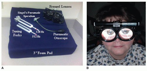
a Frenzel lenses and infrared video goggles remove visual fixation.


b Finger-to-nose, finger-nose-finger, hand rapid alternating movement test, fine finger movements, heel-to-shin, past pointing test.


c Hyperventilation-induced nystagmus may also occur.


BPPV, benign paroxysmal positional vertigo; CPA, cerebellopontine angle.


May 24, 2016 | Posted by in OTOLARYNGOLOGY | Comments Off on Clinical Evaluation of the Patient with Vertigo

Full access? Get Clinical Tree

Get Clinical Tree app for offline access