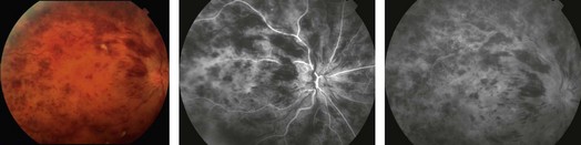
14.2 Dilated, tortuous retinal veins and intraretinal hemorrhages are noted in all four quadrants (Fig. 14.2.1). Cotton wool spots, disc edema and macular edema may also be seen. CRVO may be subdivided into ischemic or non-ischemic based on the degree of peripheral retinal non-perfusion seen on fluorescein angiography. Ischemic CRVO tend to have worse visual acuity (<20/200) and more confluent hemorrhage and cotton wool spots. Non-ischemic CRVO usually presents with better visual acuity and the relative paucity of cotton wool spots. Non-ischemic CRVO will convert to ischemic CRVO over weeks to months in about 20–30% of cases. Although the diagnosis of CRVO can be made by the characteristic fundus appearance, perfusion and the presence of ischemia is best assessed by a fluorescein angiogram.
Central Retinal Vein Occlusion
Clinical Features:
Central Retinal Vein Occlusion