Pathologic diagnosis
n
%
Melanocytic tumor
Nevus
92
47
Dysplastic nevus
2
1
Primary acquired melanosis
1
0.5
Malignant melanoma
1
0.5
Benign epithelial tumor
Papilloma
29
15
Sebaceous gland hyperplasia
15
8
Sebaceous gland adenoma
2
1
Epidermoid cyst
10
5
Oncocytoma
7
4
Sweat gland cyst
1
0.5
Pilar cyst
1
0.5
Keratoacanthoma
1
0.5
Premalignant epithelial tumor
Carcinoma in situ
2
1
Dysplasia within papilloma
1
0.5
Malignant epithelial tumor
Primary basal cell carcinoma
2
1
Sebaceous gland carcinoma
1
0.5
Lymphoid tissue tumor (low-grade lymphoma)
1
0.5
Inflammatory lesions
16
8
Miscellaneous
10
6
20.3 Epithelial Tumors
20.3.1 Papilloma
Clinical appearance typically shows a cauliflower-like mass (Fig. 20.1). Histologically, the tumors are composed of fibrovascular fronds covered by acanthotic conjunctival epithelium. There is a strong association between conjunctival papillomas and certain types of human papilloma virus, mainly types 6 and 11 [11]. Recurrence rates of conjunctival papillomas vary from 6 to 27 % [12]. Topical (one million units/cc, one drop four times daily, until clinical resolution) and/or perilesional interferon α2B (ten million units/cc injected monthly until clinical resolution) has been used to treat recalcitrant cases.
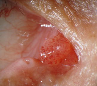
Fig. 20.1
Papilloma. A fleshy, cauliflower-like mass consisting of translucent epithelium arranged in a papillary configuration
20.3.2 Sebaceous Hyperplasia
It clinically appears as a greasy, granular, yellowish tumor (Fig. 20.2a). Histology shows mature sebaceous lobules grouped around a central duct (Fig. 20.2b).
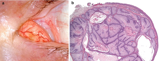
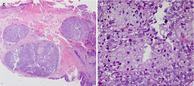


Fig. 20.2
Sebaceous hyperplasia and carcinoma. Sebaceous hyperplasia of the caruncle appears as a mound-like, yellowish subepithelial mass (a). The lesion is composed of lobules of enlarged subepithelial sebaceous glands that exhibit normal maturation. The duct of the glands may also be seen (b). Sebaceous carcinoma contains lobules of sebaceous glands that are more cellular than normal or hyperplastic glands (c). Higher magnification shows that the tumor cells in the glands contain pleomorphic nuclei with prominent nucleoli and the cells exhibit sebaceous differentiation (d) (a and b, courtesy of Ralph C. Eagle Jr., MD)
< div class='tao-gold-member'>
Only gold members can continue reading. Log In or Register to continue
Stay updated, free articles. Join our Telegram channel
Full access? Get Clinical Tree
