Fig. 10.1
Illustrations of three mechanisms of burner/stinger: (a) traction (arrows show direction of movement), (b) direct blow to Erb’s point (arrow), (c) compression (Images courtesy William H. Light Ph.D. (panel a) and Achala Talati D.O. (panels b and c))
1.
2.
3.
Compression. The third described mechanism is an extension-compression mechanism created by hyperextension of the neck and lateral bend to the affected side, effectively creating a narrowing of the intervertebral foramen and compression of the nerve root [33]. Levitz et al. implicate this mechanism as the main cause of chronic burner syndrome in 83 % of athletes, with 94 % of these athletes having associated cervical disk disease or degenerative changes [21, 27, 34].
A more recent retrospective study of Canadian football by Charbonneau et al. looked at 244 collegiate football players, noting the annual incidence of stingers to be 26 %, or 64 players in the 2010 season [26]. The most common mechanism of injury in this study was a direct blow to the shoulder. Prior to this study, the most commonly reported mechanism was the extension-compression mechanism [23, 27]. The authors postulated that this may be secondary to improved education and coaching of tackling techniques, which reduced cervical spine injuries by taking the impact to the shoulder rather than the head, but may have also placed the athlete at greater risk for brachial plexus injuries [26].
Evaluation
Initial Evaluation (On Field)
An athletic trainer responding to an athlete showing signs of distress or inability to move their arm will often conduct an initial assessment on the field. An athlete can be asked to describe what happened and what they currently feel. It is imperative to hear whether symptoms are unilateral or bilateral. Transient unilateral symptoms are less concerning, as is the absence of neck pain [35, 36]. A rudimentary strength assessment of wrist extension, biceps, deltoid, and supraspinatus can be conducted quickly with the athlete asked to resist wrist and elbow extension and resist shoulder abduction. Any deficits should prompt removal of the athlete from field of play [36–38].
Initial Evaluation (Field Side or Training Room)
In an athlete presenting with sensory symptoms in an upper extremity, it must first be determined whether the symptoms are isolated and unilateral. Any bilateral symptoms or lower extremity involvement should instead prompt suspicion for a spinal cord injury. Strength and range of motion of the neck and upper extremity should be assessed, and reflexes of the triceps, biceps, and brachioradialis should be elicited. The Spurling maneuver, in which the head is rotated to the affected side and an axial load is applied, may be positive for reproduction of radiating pain or paresthesias in athletes who have sustained an injury to the cervical nerve root, but will likely be negative in the setting of a more distal brachial plexus injury. Radiating pain reproduced by tapping on Erb’s point (Fig. 10.1, panel b) may suggest direct trauma as a mechanism of peripheral nerve injury.
Players with prompt resolution of unilateral sensory symptoms and without any weakness may be allowed to continue playing [38]. Any weakness, asymmetry of reflexes, or positive Spurling sign should prompt removal from play and further evaluation for structural cervical spine abnormalities.
Further Evaluation (Emergency Department or Office Follow-Up)
Radiographic evaluation including cervical X-ray and magnetic resonance imaging (MRI) is recommended after a first stinger if symptoms persist beyond 1 h, if there is associated neck pain, or if the neurological exam suggests a particular nerve-root distribution [20, 39]. X-ray is helpful in identifying instability on flexion or extension as well as evidence of foraminal stenosis. MRI should be performed in any athlete presenting with persistent weakness and can identify disk herniation or, as in the case of chronic burner syndrome, foraminal narrowing and disk disease [21].
Electrodiagnostic testing such as electromyography (EMG) may be helpful in evaluating the athlete with prolonged symptoms, particularly motor weakness, and is ideally performed at least 4 weeks after injury [28, 40]. Electrodiagnostics may also be helpful in distinguishing between cervical nerve root and brachial plexus injury, as well as between neurapraxic and axonal injury [41] (see Table 10.1) . This can aid in predicting time to recovery and prognosis, as neurapraxia portends a more favorable recovery [40]. A completely normal EMG is not a prerequisite for return to play, particularly if the athlete demonstrates full clinical recovery of strength and range of motion [28].
Grade 1: Neurapraxia | Grade 2: Axonotmesis | Grade 3: Neurotmesis | |
|---|---|---|---|
Severity | Mild | Moderate | Severe |
EMG findings | Focal demyelination but no axonal loss | Axonal injury resulting in positive waves and fibrillation potentials | Acute denervation |
Axon is disrupted, but epineurium is intact | Disrupts axons, endoneurium, perineurium, and epineurium | ||
Recovery | Minutes–6 weeks | Recovery may take months | Ranges from complete recovery to none |
Return to Play
Unfortunately, there is no uniform guideline for return to play after a burner or stinger; however, several practical factors s hould be considered. In general, an athlete must have full resolution of symptoms, pain-free range of motion, and normal strength [40]. If an athlete fulfills these criteria and this is their first stinger, they may return to play in the same game [29]. However, if symptoms last greater than 24 h, or there is a history of multiple stingers, then further evaluation should be considered before allowing return to play. Although it is not known how many stingers a player can safely sustain before risking permanent deficits, some experts suggest that athletes acquiring multiple stingers (e.g., more than 3) within a season should be considered for removal for the season, if not permanently [40]. Absolute contraindications to return to play include persistent weakness, persistent pain, EMG abnormalities (aside from reinnervation), reduced cervical range of motion, or evidence of myelopathy [20, 40]. Routine cervical imaging for burners or stingers in asymptomatic players is typically not recommended [29, 31, 40].
Transient Injury to the Cervical Spinal Cord
When transient neurological injury occurs to the cervical spinal cord itself, it is termed cervical cord neurapraxia . This clinical entity was described by Torg et al. in 1986 [42] and by definition involves transient functional disruption of the spinal cord from a traumatic mechanism, with subsequent complete recovery of baseline neurological function. Depending on associated motor symptoms, this has also been termed transient quadriplegia or transient quadriparesis . This injury is most commonly described in American football players , but has also been described in Rugby players [2, 43]. Affected athletes may experience numbness, tingling, or burning below the level of the injury, which either is bilateral or involves more than just the upper extremity on the affected side. Motor weakness or paralysis may also be present. Symptoms typically last several minutes but can persist up to 48 h and perhaps longer in pediatric patients [13]. A grading system based on symptom duration was developed by Torg, with Grade I injury persisting less than 15 min, Grade II 15 min to 24 h, and Grade III lasting greater than 24 h. Adults generally do not complain of associated neck pain aside from neuropathic burning pain, whereas younger children may more commonly present with neck pain [13, 14].The risk of cervical cord neurapraxia appears to increase with increasing levels of play. In a study of high school and college American football players, the incidence of cervical cord neurapraxia increased from high school to collegiate levels of play, with an annual incidence of 0.17 per 100,000 and 2.05 per 100,000, respectively [44]. Torg has previously reported an overall incidence of 7 per 10,000 elite American football players [42].
Mechanism
Penning first proposed a mechanism of injury for cervical neurapraxia in 1962, suggesting hyperextension of the cervical spine, which causes folding of the dura mater and ligamentum flavum with protrusion into the spinal canal, as well as approximation of the posterior inferior edge of a vertebral body with the anterior superior aspect of the lamina of the inferior vertebra [45–47]. This effectively causes a decrease in the diameter of the cervical spinal canal, with transient compression of the spinal cord. Hyperflexion mechanisms, theoretically with similar transient narrowing of the AP diameter of the spinal canal, have also been reported, particularly in pediatric athletes [13]. In fact, cervical cord neurapraxia is likely a mild form of SCIWORA, which is well described in children and is thought to be a result of a hypermobile cervical spine and cervical ligaments which allow movement that is sufficient to injure the cord without structural damage to the bones or surrounding connective tissues [16]. In patients with preexisting cervical spinal stenosis (congenital or acquired), the pincer mechanism may be amplified, and this has been shown to be a risk factor for recurrence [48].
Evaluation and Management
The approach to an athlete with any neurological symptoms suggestive of acute spinal cord injury should involve immediate spinal immobilization until traumatic cervical spine instability can be definitively ruled out. Plain radiographs in flexion and extension may reveal evidence of ligamentous injury or instability. Computed tomography (CT) can reveal evidence of bony injury. Any athlete with evidence of cervical cord injury should be considered for MRI, which in addition to revealing cord injury or compressive pathology (such as hematoma) can also help to establish whether there is underlying spinal stenosis which may be a predisposing factor for reinjury. Return to play decisions following an episode of cervical cord neurapraxia are an area of ongoing debate and, as discussed below, may be informed by assessment for preexisting or acquired cervical spine abnormalities.
Prevention of Cervical Neurological Injury
Modifiable Factors
Modifiable factors for prevention of both reversible and irreversible neurological injury in sport include proper equipment, proper tackling techniques, and rules modification/enforcement.
Rule changes in both American football and ice hockey have been shown to decrease the overall rate of spine injuries. The practice of spear tackling in American football, or using the helmet as the initial contact point in tackling and thereby axially loading the cervical spine in a flexed position, was banned by the National Collegiate Athletic Association (NCAA) and the National Federation of State High School Athletic Associations in the mid-1970s. During that time, a decrease in severe cervical spine injury rates was noted at both the high school and college level [49]. In ice hockey, the practice of checking from behind has been shown to result in flexion injuries to the cervical spine, and penalties for this practice were instituted in the mid-1980s in Canada [3, 5]. A decrease in spinal injuries was also noted after implementation of these rules change [5]. Quarrie et al. showed that an educational intervention via CDROM video for coaches (“Rugby Smart”) in New Zealand ultimately helped reduce the incidence of serious spinal cord injury [50].
Coaches should review proper tackling techniques, specifically avoiding the dropping of the shoulder and allowing the head and neck to be driven into extension [24]. The athlete should be able to make eye contact with their opponent in order to ensure a more vertical tackling position [24]. Postural dysfunction (stooped shoulder, head forward posture) may cause brachial plexus irritation to persist after a stinger. Chest-out posturing and thoracic outlet obstruction exercises allow the intervertebral foramina to open to its maximum [51].
Equipment modification, such as the use of thicker shoulder pads, neck rolls, or Cowboy Collars in American football, has been used to try to decrease cervical injury [52]. Such measures decrease extension and lateral flexion, as well as provide more padding for Erb’s point and thus minimize direct impact to the area, but the evidence is limited as to the efficacy of such measures for preventing stingers or cervical cord neurapraxia [52]. While no protective equipment has been definitely shown to prevent severe spinal injury in collision sports, all players should have properly fitting equipment that should be worn according to the standards in place for each sport [32].
Developmental Cervical Stenosis
Cervical stenosis has been shown by some authors to be a risk factor for permanent neurological injury in contact sports [53]. The diagnosis of cervical stenosis was previously made by measuring the canal diameter on a cervical plain film. However, this method has been shown to be subject to error due to magnification artifact, which may be present to different degrees on different films. Pavlov and Torg [54] therefore proposed evaluating the width of the canal by comparing it with the vertebral body width on the same film. This Torg ratio (or Pavlov ratio) is calculated by dividing the distance between the midpoint of the posterior aspect of the vertebral body to the nearest point on the spinolaminar line by the sagittal diameter of the vertebral body (see Fig. 10.2). In a retrospective cohort study , Torg found that a ratio of 0.8 or less was 93 % sensitive for predicting an episode of cervical cord neurapraxia [55]. However, it was found to have a poor positive predictive value of 0.2 %. Herzog subsequently showed this ratio to be a poor predictor of true functional spinal stenosis as well, with a positive predictive value of only 12 % [56]. It is now widely accepted that MRI or myelography demonstrating the degree of “functional stenosis,” or the amount of spinal fluid that is visible around the cord, is a far more accurate, though more expensive, assessment of true spinal stenosis.
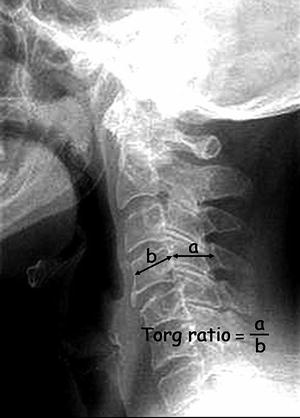
Fig. 10.2
Torg ratio. The distance between the midpoint of the posterior aspect of the vertebral body to the nearest point on the spinolaminar line (a) is divided by the sagittal diameter of the vertebral body (b)
Cervical Stenosis and Stingers
There is evidence of an association between cervical spinal stenosis and risk of sustaining a stinger injury, particularly via the nerve-root compressive mechanism. Meyer et al. demonstrated that players with cervical stenosis as assessed by the Torg ratio had a threefold higher risk of sustaining a stinger than those without stenosis [27]. In contrast, Castro et al. in their study of 165 collegiate athletes noted no difference in initial stinger frequency in players with cervical stenosis compared to players without stenosis. They did note that players with a history of repeated stingers often did have lower Torg ratios [47, 57]. Page et al. examined the utility of pre-participation screening radiographs of the cervical spine in 125 athletes and found the Torg ratio to have a positive predictive value of only 22 % for subsequent stinger experience [31]. The Torg ratio is therefore not an accurate predictor of an athlete’s risk for developing a stinger and should not be used as a screening tool [19, 31, 55].
< div class='tao-gold-member'>
Only gold members can continue reading. Log In or Register to continue
Stay updated, free articles. Join our Telegram channel

Full access? Get Clinical Tree


