(1)
University of Sydney, Sydney, Australia
Overview: The Physiology of Binocular Vision
Binocular vision is obtained from two retinal images that are fused through motor and sensory processes culminating in the perception of a single image and stereoscopic depth.
Visual information from each eye remains segregated until it passes to binocular neurons in the primary visual cortex (see Chap. 14).
Stereopsis is the sense of 3-dimensional depth perception based on slight binocular image disparity detected by these cortical neurons.
The paramount consequence of binocular vision is fine stereopsis.
This is because the following are required to achieve fine stereopsis:
(a)
Central fixation with normal visual acuity in each eye
(b)
Precise oculomotor control that achieves bifoveal fixation
(c)
Normal retinal correspondence regarding visual direction in space
(d)
The ability to perceive slight discrepancies in image location from each eye to generate a sense of depth
Binocular Single Vision
Binocular single vision (BSV) is the ability to see one image with both eyes simultaneously.
1.
Levels of binocular single vision [1–5]
(i)
Simultaneous perception
The subject simultaneously perceives an object with each eye.
(ii)
Fusion
Cortical fusion of the two retinal images leads to sensation of a single image.
(iii)
Stereopsis
Images are fused; however, slight horizontal disparity gives the perception of depth.
This is the highest level of BSV.
2.
Retinal correspondence
Corresponding retinal areas share a common subjective visual direction. When co-stimulated, these result in a sensation of single vision [6, 7].
Non-corresponding retinal areas, when co-stimulated, result in a sensation of diplopia.
Normal retinal correspondence, a cortical phenomenon, implies that corresponding areas of each retina have the same position relative to each fovea [6].
3.
The horopter and Panum’s fusion area
The points in space that project to corresponding retinal areas lie on an imaginary curved arc, the horopter, which is centered on the point of fixation (Fig. 25.1) [7, 11, 12].
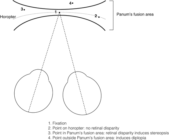
Fig. 25.1
The horopter and Panum’s fusion area
An object located on the horopter does not induce binocular image disparity.
Small amounts of disparity can be overcome by the visual system’s ability to fuse disparate images.
Hence, objects located within Panum’s fusion area (PFA), a narrow region anterior and posterior to the horopter, result in single vision [7, 10, 14].
The slight image disparity induced by objects in PFA results in the sensation of stereopsis [10, 13].
Close to fixation very little disparity is tolerated; more is tolerated farther toward the periphery [15].
Correspondingly PFA is narrow centrally and broad peripherally.
Objects outside PFA cause image disparity beyond the limits of fusion: these cause diplopia [16].
4.
Fusion
(i)
(ii)
Motor fusion
Motor fusion adjusts eye position to maintain sensory fusion.
As a fixation target approaches the observer, the retinal images move temporally from each fovea if the eye remains in an unchanged position.
To prevent diplopia, the crossed image disparity induces both eyes to converge (turn inwards) and maintain the image focused on the foveae [22].
A similar divergent movement occurs as objects move from near to far.
Fusional reserve indicates the level at which motor fusion breaks down, usually causing diplopia.
Stereopsis
Stereopsis occurs when retinal disparity is too great to permit simple superimposition of the two retinal images, but not great enough to elicit diplopia [13].
1.
Parvocellular and magnocellular stream-mediated stereopsis
Parvocellular mediated stereopsis is most sensitive for centrally located, static stimuli.
Magnocellular mediated (motion) stereopsis is most sensitive for peripherally located, moving stimuli and only capable of gross stereoacuity. It is color insensitive [33].
2.
Stereoacuity
Stereoacuity is measured as the smallest relative binocular disparity that can produce stereopsis.
Stereoacuity is greatest at central fixation and declines with eccentricity [34].
3.
Tests of stereoacuity
Several clinical tests can evaluate stereoacuity; these are designed as screening tools for distinguishing normal from abnormal binocular vision [5].
Contour stereopsis tests involve horizontal separation of the image to each eye with polarized or red-green dissociative glasses (e.g., Titmus fly test) [40].
Random dot stereopsis tests embed the stereo figures in a background of random dots e.g., TNO, Lang Stereotest (Switzerland) [41].
4.
Other mechanisms of depth perception
Stereopsis is not synonymous with depth perception: there are other clues of depth perception that are helpful for monocular individuals [42–44].
Monocular clues include object overlap, apparent size, highlights and shadows, motion parallax, and perspective.
For far (>6 m) distances, depth perception is based almost entirely on monocular clues.
Abnormalities of Binocular Single Vision
Sensory Adaptations to Strabismus
These include:
1.
Suppression
2.
Subjective Testing for Suppression and Abnormal Retinal Correspondence
They can only be interpreted in conjunction with a cover test to assess for strabismus.
BSV in the context of a manifest ocular deviation implies ARC.
These tests include:
Get Clinical Tree app for offline access
1.
Base-out prism test (Fig. 25.2)
Fig. 25.2
The 20 Δ base-out prism test
This is a test for fusion in children, used as an indirect marker of binocular vision [61].
A 20 Δ base-out prism is held in front of one eye.
If fusion is present, there will be a corrective contralateral saccade of both eyes followed by a slow refixation of the eye without the prism.
A 4 Δ prism can be used in a similar manner to detect a small scotoma in monofixation syndrome [62].
2.
Bagolini striated glasses (Fig. 25.3) [63, 64]
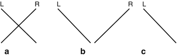
Fig. 25.3
Possible results of the Bagolini striated glasses. (a) Normal BSV. (b) Diplopia. (c) Right suppression
Each lens has striations at 90° to the other, converting a point source light to a line.
If an unbroken, symmetrical cross is seen, the patient has BSV.
If two lines that do not cross or cross asymmetrically are seen, the patient has diplopia.
If only one line is seen, or one of the lines appears broken, the patient has suppression.
Stay updated, free articles. Join our Telegram channel

Full access? Get Clinical Tree


