Amblyopia and Its Management
Scott E. Olitsky
David K. Coats
Amblyopia is derived from Greek and means “dullness of vision.” Amblyopia is a unilateral or, less commonly, bilateral reduction of best-corrected visual acuity that cannot be attributed solely to the effect of a structural abnormality of the eye or the posterior visual pathways. It is caused by abnormal visual experience as a result of patterned vision deprivation during immaturity. In most cases, it is reversible by therapeutic measures.1
Amblyopia is a functional reduction in the visual acuity of an eye caused by abnormal visual experience during the critical period of visual development. Ophthalmologic examination of the eye typically reveals no organic abnormality. The mechanism of vision loss is not fully understood, but it is thought to originate in the visual cortex. Amblyopia is the most common cause for unilateral vision loss in young people.
Epidemiology
The prevalence of amblyopia in the United States is estimated to be between 1% and 3%.2,3,4 A study of 946 children in Newfoundland found an incidence of amblyopia of 4.7%.5 Using a conservative estimate of a 2% prevalence, there are approximately 5.9 million people with amblyopia presently in the United States. Prevalence rates for amblyopia are higher in developing countries.6 The National Eye Institute has reported that amblyopia is the most common cause of unilateral visual loss in patients under the age of 70 years.7 Estimates of prevalence, however, are affected by the definition of reduced visual acuity and by the process of early screening and treatment in the population being studied.2,8,9,10 A population-based study evaluated 3,654 people age 49 and older in an area around Sydney, Australia. Amblyopia was diagnosed in 3.2% of this population, using a visual acuity criterion of 20/40 or less and 2.9% using a visual acuity criterion of 20/30 or less.11
The mean age at presentation of amblyopia varies depending on its cause.12,13 Patients with strabismus and amblyopia are more likely to present at an early age than are patients with straight eyes. In one series of children with amblyopia, the mean ages at presentation for anisometropic, strabismic, and mixed amblyopia were 5.6, 3.3, and 4.4 years, respectively.12 The upper age limit for the development of amblyopia in children who are exposed to an amblyopia-inducing condition (e.g., traumatic cataract) has been reported to be between 6 and 10 years.14 This concept is important when considering the time course for treatment of a child who sustains trauma leading to poor vision from a corneal scar or cataract.
Social and Psychosocial Factors
Detection and treatment of amblyopia is important for a variety of reasons. Affected patients may have reduced vocational and socioeconomic opportunities because normal vision in both eyes is required for many jobs, such as law enforcement, the military, and transportation work.15 Individuals with amblyopia are at increased risk for loss of vision and blindness in the nonamblyopic eye.16,17 The projected lifetime risk of vision loss in the fellow eye has been estimated to be 1.2%.16 The risk of bilateral visual impairment in patients with amblyopia is greater than twice that of patients without amblyopia.18 Amblyopia leads to a reduction in binocularity, which can lead to the development of strabismus, if not already present.
Significant psychosocial stress related to amblyopia therapy has been reported by amblyopic children and the families of amblyopic children during the treatment period.19,20 Even adults with a history of amblyopia treatment in childhood continue to have psychosocial difficulties related to the previous amblyopia therapy that can adversely affect self-image, work, education, and friendships.21
Amblyopia treatment is cost efficient.22 The care of children with poor vision secondary to amblyopia may be one of the most economically productive treatments for any vision disorder. The amount of resources and money spent per lines of vision gained for the number of years that vision will be used is significant. Membreno et al.23 utilized the concepts of time trade-off and money trade-off to compare amblyopia therapy with treatment of other medical conditions. Time trade-off involves patients reporting how many years of their remaining life expectancy would they be willing to give up to treat a given problem, whereas the money trade-off approach involves patients reporting how many months of their present income they would be willing to spend to cure a particular problem. These measures allow comparison of treatment of a diverse range of medical conditions against each other. These authors reported on the incremental cost-effectiveness of therapy for amblyopia and calculated a savings of $2,281 per quality-adjusted life year with amblyopia treatment. They concluded that when compared with health care interventions for other medical conditions, amblyopia care is highly cost-effective.23
Visual Development
During fetal development, differentiation and organization of the visual system are likely guided by intrinsic control mechanisms. At birth, the process is not complete and must continue to develop throughout the first decade of life. Unlike the prenatal period, environmental factors and visual experience influence the process in postnatal life. For normal visual development to occur, both eyes must be presented with equally clear and similar images.24 This constant stimulation is crucial to the development of normal vision. Any process that interferes with this process can lead to the development of amblyopia.25 The human visual system is sensitive to the effects of depriving vision only during a limited period of time in childhood. This time period is often called the sensitive or critical period. For humans, this period extends roughly from birth through the end of the first decade of life.14,26 Vulnerability is greatest during the first few months of life and thereafter gradually decreases.
Abnormal visual experience during the critical period can take one of two forms.27 In the first, a lack of exposure to the well-formed images necessary for normal development interferes with the maturation of form vision. In the second, a marked disparity in the quality or directionality of inputs from the two eyes leads to abnormal competitive binocular interaction. This competition leads to active interference with, or exclusion of, one eye’s input to higher visual centers that persists during monocular viewing. Either of these two mechanisms can contribute to unilateral or bilateral amblyopia.
Although amblyopia often develops as a result of abnormal binocular interaction, it is not truly a disorder of binocular vision. The pathophysiology of amblyopia differs from strabismic suppression and anomalous retinal correspondence (ARC), although they are both frequently associated consequences of abnormal visual experience and can occur together in the same individual. Suppression and ARC are not defects, but are more appropriately considered adaptations of binocular vision. Unlike, amblyopia, they can benefit the patient by eliminating diplopia without undermining the capacity for normal visual function. Amblyopia has no value for the patient (diplopia can occur even when one eye is severely amblyopic), and the patient with amblyopia cannot return to a state of normal vision quickly by closing one eye or normalizing the eyes’ alignment.
Classification of Amblyopia
Amblyopia is often defined as a difference in visual acuity of two lines or more (Snellen or equivalent) in a child with an otherwise healthy visual system. In reality, amblyopia may be present any time visual acuity is reduced even if the difference is one line or less. Amblyopia can also occur in association with organic defects of the eye or elsewhere in the visual system and will be discussed later in this chapter. In addition, amblyopia leads to changes in vision that may not be detected on a Snellen acuity chart. Amblyopia is most commonly characterized by clinical associations that initiate the problem. Therefore, amblyopia is most often classified based on the causal mechanism and evidence from laboratory investigations confirms that different pathophysiologic disturbances underlie the occurrence of amblyopia in these different clinical settings. Familiarity with this classification system is important for clinicians and can be useful in designing and implementing appropriate treatment strategies.
Strabismic Amblyopia
Strabismic amblyopia is one of the most common forms of amblyopia. It results from abnormal binocular interaction that occurs when the visual axes of the two eyes are misaligned and the patient develops a preference for one eye. Amblyopia generally does not develop if the patient alternates fixation. This abnormal interaction causes the foveae of the two eyes to be presented with different images. Disparate input from the deviating eye stimulates active inhibition of the retinostriate pathways of visual projections originating in the fovea of the deviating eye. A decrease in these projections can then lead to the development of amblyopia.
Any type of strabismus can be associated with amblyopia. As many as 17% to 40% of children with congenital esotropia develop amblyopia.28,29 Patients with exotropia may also develop amblyopia. The development of amblyopia in intermittent exotropia occurs less frequently than it does in esotropia, most likely because of the intermittent nature of the disorder as well as the older age group in which it occurs. Paralytic strabismus may or may not be associated with amblyopia, depending on severity of the defect and the child’s ability to maintain fusion by adopting an anomalous head posture. Amblyopia is unusual in congenital superior oblique paresis because affected children usually adopt an anomalous head tilt to the contralateral side of the affected muscle, thus providing protection against amblyopia.
Surgery to correct the strabismus is generally considered ineffective in the treatment of associated strabismic amblyopia. The success rate of surgery for esotropia has been reported to be unaltered by the presence of mild residual amblyopia.30 Although Lam et al.31 demonstrated that surgery can treat amblyopia in some patients with esotropia, strabismus surgery is generally deferred until the amblyopia has been maximally treated. This practice allows easier detection of fixation preference in preverbal children and helps to encourage parents to comply with the sometimes difficult task of amblyopia therapy.
Anisometropic Amblyopia
Anisometropia is another frequent cause of amblyopia. Anisometropic amblyopia may occur in children with hyperopia, myopia, or astigmatism. Anisometropic amblyopia tends to occur more commonly and to a worse degree in children with anisohyperopia. This occurs because the fovea of the more ametropic eye in a child with anisohyperopia never receives a clearly focused image when viewing binocularly. In mild to moderate anisomyopia, the more myopic eye can be used for near work and the less myopic eye can be used for distance work providing an important measure of protection against development of amblyopia.
Weakley and Birch32 studied refractive errors likely to produce amblyopia. They noted that anisohyperopia as small as 1.0 diopters (D), anisomyopia as small as 2 D, or anisoastigmatism as small as 1.5 D was sufficient to produce amblyopia. Although there is significant latitude to decide when and how to treat an individual patient with anisometropia, these are excellent suggestions. Published guidelines for the prescription of spectacles in children closely resemble these suggestions.33
In contrast to the other types of amblyopia, anisometropic amblyopia may not be detected until the child is old enough to undergo vision screening performed by a pediatrician or school system. The typical child with anisometropic amblyopia lacks obvious external abnormalities of the eyes (e.g., cataracts, strabismus), and visual function appears normal because the child sees well with the fellow eye. In these patients, photoscreening may be especially useful in detecting amblyopia earlier than may otherwise be possible. Bilateral myopic shift during late childhood or adolescence may account for the occasional finding of amblyopia in the emmetropic eye of an adult with unilateral myopia, which during early childhood was more hyperopic than its fellow eye.
The mechanism responsible for the development of amblyopia in patients with anisometropia is thought to be similar to that which occurs in those with strabismic amblyopia. Active cortical inhibition of input from the fovea of one eye occurs to eliminate sensory misperceptions caused by having a focused image in one eye and a defocused image in the other.
Ametropic Amblyopia
Severe symmetric refractive error (isoametropia) can cause bilateral amblyopia of mild to moderate degree. Hyperopia in excess of about 5 to 6 D is usually needed to cause this form of amblyopia. Myopia, unless extremely high, usually does not cause bilateral amblyopia because the sharply focused images of objects held close to the eyes support normal visual development. Bilateral amblyopia is less likely to develop in the high hyperopic patient when accommodative esotropia is present. In such cases, the accommodation that causes excessive convergence has the beneficial effect of bringing the fixating eye’s retinal image into focus, whereas the nonesotropic, highly hyperopic person may not be accommodating sufficiently to have a clear view with either eye. High degrees of astigmatism can also lead to the development of ametropic amblyopia. A person who did not receive optical correction for severe astigmatic refractive error in childhood sometimes shows persistent impairment of corrected vision that is confined to the more ametropic meridians. This phenomenon is known as meridional amblyopia. It can be unilateral or bilateral. The effect on conventionally measured acuity is generally small. Ametropic amblyopia and symmetric levels of myopia may be detected in patients early because of the poor vision owing to the high levels of the myopia itself. Patients with high degrees of hyperopia or astigmatism may show less reduction in their vision and therefore the condition may not be diagnosed until they undergo some form of screening or testing.
Visual-Deprivation Amblyopia
Deprivation amblyopia is the least common and most serious form of amblyopia. Visual deprivation is caused by occlusion of the visual axis. Congenital cataracts, ptosis, congenital corneal opacities, and vitreous hemorrhage can lead to deprivation amblyopia. Even temporary obstruction of the visual axis, such as that caused by a hyphema, or temporary eyelid edema in a very young child can produce visual-deprivation amblyopia. Visual-deprivation amblyopia can be unilateral or bilateral. Amblyopia is more likely to occur, be more severe, and be more resistant to treatment when the defect promoting its development is unilateral. Sensory strabismus often occurs in children with unilateral vision deprivation. Deprivation amblyopia can result in permanent visual impairment if it is not treated urgently in infancy. Children with unilateral congenital cataracts require rapid visual rehabilitation to provide the best chance for good vision. In one series of patients with unilateral congenital cataracts, no patient achieved vision better than 20/50 who underwent surgery after 17 weeks of age.34
Special Forms of Amblyopia
Occlusion amblyopia is an iatrogenic condition caused by therapeutic patching of the eye with normal acuity. It usually occurs in the sound eye as a result of amblyopia treatment, but it is occasionally seen in young children after occlusion therapy to treat ocular pathology, such as a corneal abrasion. Concern for occlusion amblyopia is one of the most important reasons for routine follow-up of children being treated for amblyopia. Rapid development of occlusion amblyopia in the sound eye during therapy for amblyopia is a sign of continued visual system plasticity, and it is believed that it often portends a satisfactory visual outcome for both eyes of such patients if detected and corrected promptly.35
Idiopathic amblyopia is occasionally diagnosed in retrospect when a child with a monocular reduction of visual acuity and no detectable cause responds with improved vision during a trial of treatment for amblyopia. Therefore, amblyopia treatment is often attempted when visual acuity is reduced and no cause can be found. Presumably, an amblyopiogenic process was present earlier in the child’s life that has since resolved.36,37 A small angle strabismus may be discovered once the vision increases and fixation has improved. Detailed history taking may identify a history of strabismus or previous occlusion of the visual axis, for example, from prolonged eyelid edema caused by infection of the lids as a young child. Equalization of a previous anisometropic refractive error is another possible explanation and the history is unlikely to be helpful in this situation.
Organic Amblyopia
Although amblyopia generally occurs in an otherwise normal eye, it can sometimes be superimposed on visual loss directly caused by a structural abnormality of the eye, such as optic nerve hypoplasia, coloboma, or a partial cataract.38 When such a situation (“organic amblyopia”) is encountered in a young child, it is appropriate to undertake a trial of occlusion therapy. Patching may be both diagnostic and therapeutic in doubtful or borderline cases. Improvement in vision confirms that amblyopia was indeed present.
Vision in Amblyopia
Although it is most often detected, and even defined by, demonstrating a reduction in Snellen visual acuity, amblyopia leads to other defects in vision. The full range of abnormalities present in the amblyopic eye is probably not yet fully understood. They may have important clinical applications. Some of these abnormalities may be useful in detecting amblyopia or determining its cause.
Visual Acuity
Patients who are amblyopic are deficient in their ability to resolve closely spaced contours and recognize the patterns they form. Visual acuity measured with conventional tests, which rely on both these functions, will therefore be subnormal in patients with amblyopia. Measurement of visual acuity is therefore the primary manner in which amblyopia is detected. Children old enough to identify letters are usually tested with standard letter optotypes, either projected, wall mounted, or computer generated. For younger children, testing may be modified to permit the use of manual pointing responses.
Currently, the nonverbal Snellen equivalents most widely used in North America are the tumbling E test and the HOTV test. For most 3-year-olds and nearly all developmentally normal 4-year-olds, fairly reliable visual acuity measurements can be obtained with either of these tests after brief instruction (and confirmation of the patient’s competence in responding) using large demonstration letters. An advantage of the HOTV test is that there is no need to discriminate left or right mirror image figures, which may be difficult for the young patient.
Various acuity tests have been devised that substitute pictures for letters. These include Allen pictures and Lea symbols. Lea symbols consist of four figures (Fig. 10.1) that are easy for most toddlers to recognize and are more similar in configuration to Snellen letters than the Allen pictures. They have been carefully calibrated and assessed for reliability.39 Some small children who will not point to identify letters may respond to testing with these figures. Lack of familiarity with the pictured objects or inability to recognize the stylized images may be a problem for some children; testing is often begun with a review of the cards up close, and any that are not readily identified should be eliminated (three or four remaining different pictures are sufficient for testing). Clues derived from the overall shape of the Allen images may undermine the accuracy of acuity measured. In general, other recognition tests noted above are preferred over Allen figures, when possible. On the occasion when a switch is made from Allen pictures to letters (or any significant change in the method of acuity assessment is made), it may be helpful to record the results from both tests to help ensure comparability of measurements over time.
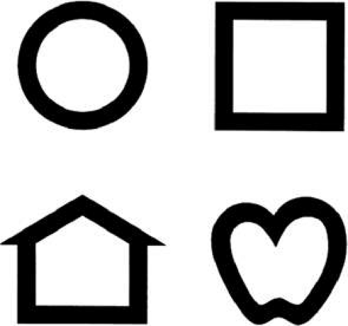 Figure 10.1 The four Lea symbols |
Accommodation
The function of accommodation is to bring retinal images into sharp focus, a state with which the amblyopic eye often lacks experience and which it may have difficulty recognizing. It is not surprising that amblyopia impairs the ability to control accommodation, typically resulting in a subnormal response.40,41,42 This subnormal response may occur because there is no stimulus to elicit an accommodative effort. If the image is brought into focus, it will not appear clearer because of the underlying amblyopia. In essence, the effort of accommodation is not rewarded by providing the patient with improved vision. In addition, amblyopic eyes have a decrease in the ability to accommodate as well and demonstrate a decrease in accommodative amplitude. Therefore, moderate hyperopia that would not be expected to affect the vision of a normal eye, which will accommodate to sharpen its retinal image, may cause significant optic blur in association with amblyopia.
The effect of amblyopia on accommodation should be considered when prescribing glasses in patients with reduced vision. Leaving a portion of a hyperopic refractive error uncorrected, may alter the course of amblyopia treatment in some patients. For this reason, that some practioners may prescribe the full amount of hyperopia found during a cycloplegic refraction to patients with amblyopia.
Spacial Interactions
Patients with amblyopia have increased difficulty identifying test letters when they are presented in a linear or two-dimensional array compared with those presented as isolated characters. This “crowding phenomenon” is an example of the effect of contour interaction on visual acuity. It can be demonstrated in normal and organically diseased as well as in amblyopic eyes when figures near the limit of resolution are surrounded by other closely spaced forms. In the normal fovea, contour interaction occurs when forms are separated by a distance of 1 to 3 minutes of arc (0.2 to 0.6 times the overall size of a 20-foot [6-M] Snellen letter). In the amblyopic fovea, contour interaction typically extends over an increased distance, to a degree that is roughly proportional to the reduction in acuity.43
Other conditions that decrease visual acuity may produce similar extension of contour interaction.44 The magnitude of the effect on measured acuity in amblyopic eyes is unique and may result from this phenomenon. For example, occasionally, an amblyopic eye that can resolve a 20/20 letter in isolation drops to as low as 20/100 in the presence of maximal contour interaction. These larger discrepancies sometimes develop in the course of treatment as acuity measured with isolated letters improves more rapidly than interactive acuity.45 Therefore, amblyopia should not be considered fully treated until interactive acuity has been maximized. This may pose a dilemma when testing vision in young children. It may be difficult to test younger patients with linear figures and isolated figures may make milder cases of amblyopia more difficult to detect. The crowding phenomenon can be induced through the use of interactive bars around a single letter or figure (Fig. 10.2). Red crowding bars that surround black optotypes appear to create a greater effect than the use of black bars.46
Stay updated, free articles. Join our Telegram channel

Full access? Get Clinical Tree


