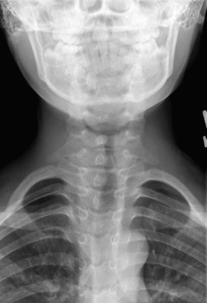
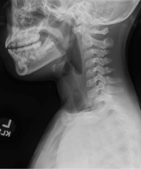
67 A 7-year-old girl was brought to the emergency department for escalating stridor. She was initially taken to her pediatrician 2 weeks ago for noisy breathing, where she was diagnosed with asthma and given an albuterol inhaler. Despite albuterol therapy, her breathing became progressively labored. On further questioning, her mother relates the onset of stridor to a brief coughing and choking spell after the patient had been playing with a toy tea set. On examination in the emergency department, the patient demonstrated biphasic stridor. Her respirations were regular with equal breath sounds, and her skin color was pink. She became uncomfortable when placed supine, but she was in no acute distress. Posterolateral and lateral neck radiographs were obtained that showed a slightly opaque, geometrically shaped foreign body at the level of the larynx (Figs. 67.1 and 67.2). 1. The most common causes of stridor in an infant are laryngomalacia, vocal cord paralysis, and subglottic stenosis. These congenital causes of stridor should have manifested themselves within the first year of life. Subglottic hemangiomas also cause biphasic stridor, but the onset is gradual; symptoms reach their maximum around 8 to 12 months. 2. Infectious and inflammatory processes frequently affect the pediatric airway. A prodromic upper respiratory illness that leads to stridor may indicate croup or bacterial tracheitis. More rarely, rapid-onset stridor in a toxic child is consistent with epiglottitis. 3. Foreign bodies must always be considered in the pediatric population (typically 10–24 months of age), and frequently foreign body ingestion is an unwitnessed event. Stridor is common in fixed obstructions of the larynx or trachea. Chronic esophageal foreign bodies may also cause airway compression and secondary tracheomalacia by irritating the common wall between the trachea and esophagus. The mortality rate for foreign bodies in the upper aerodigestive tract is around 2%, with laryngeal foreign bodies having a mortality rate approaching 45%. Physical examination must assess the patient’s respiratory rate, color, and degree of distress. Listening to the breath sounds will inform the location of the airway foreign body. Classically inspiratory stridor is consistent with supraglottic pathology, but a large supraglottic foreign body will also induce gagging, retching, and a muffled voice. Laryngeal foreign bodies demonstrate biphasic stridor, whereas foreign bodies in the intrathoracic trachea cause expiratory stridor. Chest auscultation may indicate ispilateral wheezing, cough, and decreased breath sounds, but this will occur in less than 50% of the cases. A prolonged inspiratory or expiratory phase of respiration is also consistent with a distal airway foreign body. The diagnostic yield from the physical examination improves with chronicity. Fig. 67.1 Anteroposterior neck radiograph demonstrating a radiopaque geometric foreign body in the subglottis. In a stable patient, the initial evaluation of the airway is a high-kilovolt posteroanterior lateral neck and chest radiograph. The structural elements of the airway are outlined, and radiopaque foreign bodies may be identified. The lung fields can also be evaluated for signs of postobstructive emphysema. If a foreign body is ball-valving into a bronchus, there could be air trapping and hyperinflation of the lung field on that side, and mediastinal structures will shift away from the obstruction. Conversely, a completely obstructed bronchus may lead to absorption of the air trapped distally with widespread atelectasis. In these cases, the mediastinum will shift toward the obstruction. Fig. 67.2 Lateral neck radiograph demonstrating a radiopaque geometric foreign body in the subglottis.
Airway Foreign Body
History
Differential Diagnosis—Key Points
Test Interpretation
Stay updated, free articles. Join our Telegram channel

Full access? Get Clinical Tree


