Surgical navigation technology provides real-time intraoperative localization of surgical instruments within the field. These systems are highly accurate, assist with preoperative planning, and improve surgeon confidence. The industry has recently responded to the growing trend of treatment in ambulatory surgical centers by offering surgical navigation devices that are more compact, less expensive and more user-friendly than conventional devices. Surgical navigation is indicated for complex sinonasal disease and may reduce the risk of complications. The indications for surgical navigation continue to expand as the technology improves and imaging data synthesis evolves to include multimodality fusion and real-time intraoperative data-set updates. Although now widely available, navigation systems are still considered state of the art, and not standard of care.
Endoscopic sinus surgery using rigid endoscopes is now the conventional technique for opening obstructed and diseased sinus outflow tracts. Although the application of endoscopes has revolutionized the diagnosis and surgical management of sinonasal disorders, a major limitation continues to be that the view is not in three dimensions. When operating from only a two-dimensional magnified image, localization of instruments within the sinonasal cavity during surgery depends largely upon the depth of penetration and the quality of tactile feedback. Additionally, surgical orientation may be limited by distorted anatomy, extensive polyposis, or intraoperative bleeding. Navigation technology may be viewed as a method to improve visualization during surgery and serves as an adjunct to nasal endoscopy. Surgical navigation systems can be employed to determine the precise location of critical structures during the course of an operation.
Navigation systems identify anatomic landmarks by locating surgical instruments in space, calculating the location of the instrument tip in relation to the patient, and projecting the instrument location onto a previously obtained imaging study (usually computed tomography [CT]). The operator can use this information for intraoperative surgical navigation or preoperative planning using the computer workstation, which displays the patient’s images simultaneously in all three anatomic planes (coronal, axial, and sagittal).
Surgical navigation components
Imaging Data
CT scans are the imaging modality of choice when evaluating the paranasal sinuses and are used routinely for image-guided surgery. The detailed bony anatomy provided by high-resolution CT imaging accentuates the surgical landmarks and the confines of the sinonasal cavity. The unit of data in CT imaging is cuboidal and permits reconstruction of images in the coronal, axial, or sagittal plane. Thinner slices improve resolution. The optimal CT scan is an axial CT with 1-mm slice thickness and a 512 × 512-pixel matrix.
Magnetic Resonance Imaging (MRI) provides excellent soft tissue detail and is frequently incorporated into image-guidance systems for intracranial procedures. MRI is also extremely useful in the evaluation of sinonasal conditions, such as allergic fungal sinusitis and neoplasia, precisely detailing the site and extension of benign and malignant sinonasal tumors. MRI is also useful in evaluating encephaloceles and cerebrospinal fluid leaks, as the high-intensity signal of cerebrospinal fluid in the sinuses on T2-weighted imaging can suggest the presence of a leak. Like CT data, MRI data can be loaded into an image-guidance system and used for intraoperative navigation.
Traditionally, the imaging data are obtained preoperatively and transferred to the image-guidance system computer through digital storage media, such as universal serial bus (USB) sticks or compact disk–read-only memory (CD-ROM) disks. Newer systems have the capacity to directly download data via high-speed Internet or wireless Ethernet connections, making data transfer from radiology to the navigation system in the operating room simple and seamless. Until recently, most available image-guidance systems required a special proprietary CT protocol for the data set to be incorporated into the software and CT scans performed elsewhere (referred to as foreign data sets) were not usable for navigation. This was a major shortcoming, as it mandated repeat imaging, thus exposing the patient to further radiation, adding to inconvenience, and increasing cost, solely for the purpose of employing navigation. Manufacturers of most systems have resolved this issue such that the majority of foreign data sets available in electronic format are now suitable for navigation provided they conform to a few common parameters. These vary somewhat by manufacturer but frequently include axial cuts with a 512×512-pixel matrix, 3-mm or less slice thickness, and a 0° gantry tilt. This has been a significant improvement in software design for these systems, and has made navigation easier to use.
Registration
Registration is the establishment of a rigid relationship between two coordinate systems: (1) the images previously obtained and (2) an arbitrary system employed to describe every point within the surgical field. After registration is performed, a navigation system is capable of translating from any point on the patient to the same point on the imaging study using a transformation matrix.
Three types of registration are currently available: paired-point registration, automatic registration, and contour-based registration. The paired-point method requires the surgeon to designate fiducial points on both the patient and imaging data. Accuracy can be improved by increasing the number of points and using a distribution of fiducial points to encompass the three-dimensional area of surgical interest. A greater number of paired points is desirable; however, a greater number of points increases the time for registration and does not significantly improve accuracy. Most surgeons recommend 6 to 10 paired points in a nonlinear distribution around the surgical area. Automatic registration requires a headset that is placed on the patient at the time of CT or MRI. The headset, embedded with metal fiducials, is designed to fit the patient in a reproducible fashion and it must be reapplied and worn during surgery. The image-guidance system recognizes points within the headset and uses these for “automatic” registration. The accuracy of this method depends on consistent placement of the headset. Contour-based registration requires the image-guidance system to build a three-dimensional model of surface contours of the patient’s face using the imaging data. A standard probe or reflected laser light can then be used to define the surface contour of the patient at the time of surgery. This technique rapidly defines a large number of registration points (40–500). The contour method loses accuracy if the patient’s surface contour changes from the time of image collection to the time of surgery because of edema, weight gain, and stretching from surgical drapes. Early registration paradigms were difficult to follow, but recent features, such as touch screens, auto-registration, and surface registration, have simplified the process significantly. Most systems now employ either automatic or surface registration given the greater simplicity of these techniques. Multiple clinical trials have shown all three registration systems to be reliable with accuracies of 1 to 2 mm.
Tracking Technology
Tracking is a method of dynamically following the movement of an instrument in space, calculating the location of the instrument in relation to the patient, and projecting the location of the instrument onto an imaging study. The two tracking systems currently available are optical- and electromagnetic-based systems. Both systems are highly accurate to within a few millimeters and both systems account for intraoperative head position by fastening a headset to the patient.
Electromagnetic systems track instruments using a radiofrequency transmitter within a special headset and a receiver positioned within the instrument. The headset, containing embedded fiducial markers, and trackable instruments are attached via wires to the system hardware. The headset must be worn during the preoperative CT scan and again intraoperatively. Electromagnetic tracking may be susceptible to interference from metal instruments within the field or the metal operating room table. Thus, metal devices and anesthesia equipment must be placed an appropriate distance away from the operating field and some investigators recommend placing extra padding between the patient and the table.
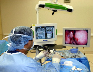
Optical systems use infrared light and a camera positioned 6 ft (approximately 1.8 m) above the patient’s head to track instruments ( Fig. 1 ). This provides a wireless mechanism for instrument localization, but requires a clear pathway between the tracking device and the instruments. Equipment and draping must be placed carefully to avoid line-of-sight obstruction.
Specialized equipment and instruments are needed for both types of systems. Optical tracking systems require light-reflecting spheres (also referred to as stars or glions ) attached to the proximal end of wireless surgical instruments. Electromagnetic platforms require the attachment of a wired probe to the instruments. Both systems allow for a variety of instruments to be tracked, including probes, straight and angled suctions, and curettes. With the optical-based systems, however, a recent innovation known as universal calibration allows reflective spheres to be affixed to any rigid instrument, thus permitting navigation with any surgical instrument including drills and microdebriders. The computer workstation integrates the data set with information from the tracking system and projects the location of the instrument tip.
During the course of surgery, the surgeon must frequently corroborate the accuracy of the image-guidance system using obvious surface and intranasal landmarks, such as the tip of the nose or the posterior wall of the maxillary sinus. A change in the angle of the instrument facing an optical tracking device or movement of the headset can change the projected location and contribute to inaccuracies and drift.
Surgical navigation indications
In general, the indications for surgical navigation are situations in which additional information beyond the endoscopic or surgical view is beneficial. This includes situations involving patients with advanced disease or unusual pathology. Patients undergoing intervention for disease in close proximity to critical structures, such as the skull base or orbit, or in cases where the disease extends to difficult-to-reach anatomic locations (perhaps even beyond the paranasal sinuses) would benefit from surgical navigation. The American Academy of Otolaryngology has endorsed the use of image-guidance and has described examples of indications for navigation ( Box 1 ). In addition to the intraoperative advantage with image-guidance, the display of the imaging data in three simultaneous planes can be a very powerful tool for preoperative planning, particularly important in more complex cases. Surgical navigation may also improve resident training by demonstrating anatomic relationships and allowing for monitoring of resident progress.
- ○
Revision sinus surgery
- ○
Distorted sinus anatomy of development, postoperative, or traumatic origin
- ○
Extensive sinonasal polyposis
- ○
Pathology involving the frontal, posterior ethmoid, and sphenoid sinuses
- ○
Disease abutting the skull base, orbit, optic nerve, or carotid artery
- ○
Cerebrospinal fluid rhinorrhea or conditions where there is a skull-base defect
- ○
Benign and malignant sinonasal neoplasms
Surgical navigation is helpful, but not necessary, in patients with intermediate disease. Examples include patients with obstruction of the frontal or sphenoid outflow tract. However, an image-guidance system can be invaluable for procedures, involving revision surgery on the frontal and sphenoid sinuses.
Surgical navigation is not necessary in patients with disease localized to the maxillary and anterior ethmoid sinuses. Furthermore, the use of image-guidance should be left to the sole discretion of the individual surgeon. Surgical navigation may not be needed for any given procedure if the experienced surgeon is comfortable performing the operation with traditional techniques. Many endoscopic procedures can be performed safely with standard equipment. Therefore, use of image guidance is not standard of care.
Stay updated, free articles. Join our Telegram channel

Full access? Get Clinical Tree


