Wound construction

CHAPTER 11 Wound construction


See Video image


Correct knowledge and implementation of wound architecture are paramount in creating a stable operating environment and a safe postoperative recovery.


This chapter will cover extracapsular (ECCE), small incision cataract surgery (SICS) wounds and phacoemulsification wounds. The vast majority of cataract operations in the developed world are performed via a phaco wound but occasionally it may be necessary to perform a larger wound. SICS is, at the time of writing, becoming very popular in developing countries due to its relative similarity to ECCE, low cost, and effectivity. It would be comforting to know that long-term data in terms of endothelial cell loss and glaucoma resulting from SICS will be collected and analysed but, due to the nature of the places where SICS is mainly performed around the world and the facilities available, this is unlikely.


ECCE


A backward sloping corneal wound or stepped scleral wound may be created to allow the wound to be self-sealing and allow the use of sutures which will stop the wound from opening rather than keeping it closed. This allows the tension in the sutures to be adjusted in such a way that minimal induced astigmatism is caused.


The wound will also heal at this predecided tension, which means the sutures can be left in for around 4 months, which mitigates against wound slip from early removal. The wound should be made just big enough to express the nucleus without undue force and this means an accurate preoperative assessment of the nuclear density to allow such a judgment on wound size to be made.


Scleral ECCE wound


The architecture of the wound is similar between scleral and corneal ECCE wounds but, for a scleral wound, the conjunctiva must be reflected first and sutured back in place at the end. If a very peripheral corneal wound is made then consideration should be given to moving the conjunctiva as well because if this is not done the closing sutures which start in the cornea will exit through the sclera and conjunctiva and will ‘cheese-wire’ or cut through the conjunctiva leading to inflammation, sterile conjunctival infiltrates, and loosening of the sutures.


The wound is sloped backwards meaning that the blade used for the incision is pointing towards the surgeon. This makes the wound self-sealing and stable. If the wound is forward sloping it will leak and make the operation difficult and the postoperative period more risky.


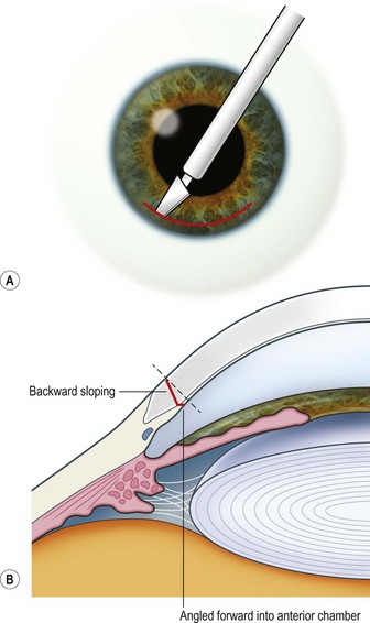
The scleral wound is made around 120°, initially backward sloping and then, when the deeper tissues are reached, a forward entry into the eye is made (Fig. 11.1). More bleeding is likely because of perforating scleral vessels and diathermy is a prerequisite. Careful wound construction and suturing can be rewarded by very little surgically induced astigmatism because of the distance from the optical centre of the cornea and the ability to leave the sutures at the desired tension.


Excessive cautery may shrink the edges of the wound and make wound apposition more difficult when closure is attempted.


Corneal ECCE wound


This should be made approximately 1 mm into clear cornea and sloped gently backwards. This ensures that the sutures when placed to close the wound will enter and exit corneal tissue and avoid the need for reflecting the conjunctiva (see Fig. 11.1).


The wound is made at least 90% depth and then a final, forward entry into the anterior chamber is made. A diamond bladed knife is best for this but a sharp steel blade is adequate, more readily available and much cheaper. Re-used steel blades blunt very quickly and should not be relied on as soon as their lack of sharpness is perceived.


The relative advantages and disadvantages of scleral and corneal ECCE wounds are summed up in Table 11.1.


Table 11.1 Comparison of corneal and scleral section


Only gold members can continue reading. Log In or Register to continue

Stay updated, free articles. Join our Telegram channel

Jun 4, 2016 | Posted by in OPHTHALMOLOGY | Comments Off on Wound construction

Full access? Get Clinical Tree

Get Clinical Tree app for offline access
Section