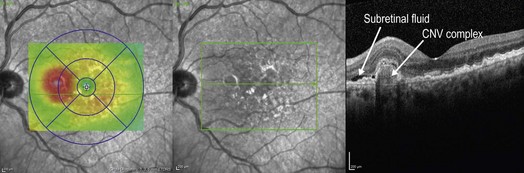
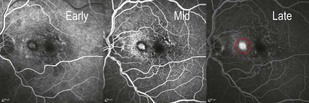
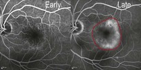
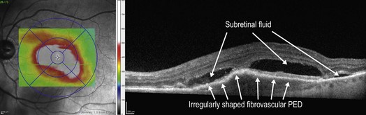
8.1 ▶ Classic CNV: a classic, or type 2, CNV is present when the abnormal neovascular tissue penetrates the RPE/Bruch’s membrane complex and is present in the subretinal space (Figs 8.1.1 and 8.1.2). Figure 8.1.1 OCT of a classic choroidal neovascularization (far right). Corresponding thickness map (left) and infrared image (middle) are shown. Figure 8.1.2 Fluorescein angiography (corresponding to Figure 8.1.1) shows a well-defined region of hyperfluorescence that is visible in the early frames and grows in intensity in the late frames, but does not enlarge in size, characteristic of a classic choroidal neovascularization (red circle). ▶ Occult CNV: an occult, or type 1, CNV is present when the abnormal neovascular tissue remains underneath the RPE (Figs 8.1.3 and 8.1.4). Figure 8.1.3 Fluorescein angiography shows late hyperfluorescence with ill-defined boundaries, which may represent a fibrovascular pigment epithelial detachment, and is characteristic of an occult choroidal neovascularization (within red border). Figure 8.1.4 OCT (corresponding to Figure 8.1.3) of an occult choroidal neovascularization (right) and corresponding thickness map (left), PED.
Wet Age-Related Macular Degeneration
OCT Features:
![]()
Stay updated, free articles. Join our Telegram channel

Full access? Get Clinical Tree


Wet Age-Related Macular Degeneration
