Fig. 11.1
Human newborn (a), infant (b), child (c), and adult (d) macula flavae and growth and development of the human vocal fold mucosa. TC thyroid cartilage, ACT anterior commissure tendon, AMF anterior macula flava, PMF posterior macula flava, P vocal process of arytenoid cartilage, RS Reinke’s space, VL vocal ligament, LP lamina propria of the vocal fold mucosa (Reproduced from Sato et al. [12])
Ishii et al. visualized collagen fibers and elastic fibers separately in pediatric cadaveric vocal folds by scanning electron microscopy after digestion treatment with sodium hydroxide and formic acid and noted the development of fibrous protein organization in vocal fold lamina propria by 17 years of age [15] (Fig. 11.2). They reported that the vocal fold lamina propria in fetuses and neonates consisted of sparse and relatively dense areas of collagen and elastic fibers that ran at random and no longitudinal arrangement was found, and the vocal ligament was not found. In subjects 5 years of age, a deep dense area was found in the anterior and posterior maculae flavae, and longitudinal fibers were noted between the maculae. A structure of superficial versus deep layers appeared in children older than 10 years of age. The layered structure of the lamina propria was complete around 17 years of age [15] (Fig. 11.2).

Fig. 11.2
Developmental changes in lamina propria of human vocal fold. Asterisk anterior macula flava, # posterior macula flava, star vestige of deep part, d dense area, s sparse area, l longitudinal fibers, lcc longitudinal curly collagen fibers, le linear elastic fibers (Reproduced from Ishii et al. [15])
Nita et al. studied the fine structure of the vocal folds from human fetuses and neonates and analyzed collagenous and elastic fibers in the lamina propria by using light and electron microscopy [16]. Collagen fibers were viewed using the Picrosirius polarization method and elastic system fibers were stained using Weigert’s resorcin–fuchsin after oxidation with oxone. They reported that the distribution of collagen and elastic fibers in the lamina propria of fetal vocal fold resembled that previously described for the adult vocal ligament, suggesting that a vocal ligament has already begun to develop by the time of birth [16].
Thus, there have been discrepancies regarding the composition and orientation of collagen and elastin fibers in pediatric vocal fold lamina propria. It is probably because the methods for visualizing the fibrous proteins used in these reports differed in sensitivity and specification. There seems to be no consensus as to when the vocal ligament first emerges so far. Accumulating evidences indicate that extracellular matrix components such as collagen and elastin are present in vocal fold lamina propria from the fetal period, leaving the question whether such fibrous proteins form the structure that can be called as vocal ligament. It is widely accepted that the organization of fibrous proteins undergoes considerable development after birth until it attains the highly complex structure seen in adults.
11.3 Development of Macula Flavae in Pediatric Vocal Folds
The human vocal fold is known to have macula flavae in both the anterior and posterior ends of its membranous portions and the vocal ligament runs between the anterior and posterior macula flavae, as described in Chap. 13.
In the fetal period, macula flavae in human vocal folds develop asynchronically with the posterior macula flavae appearing before the anterior ones [17]. The earliest appearance of posterior macular flavae have been observed at 11 weeks of amenorrhea [17], 13 weeks of amenorrhea [18] or 13.5 weeks of amenorrhea [19], whereas the anterior macula flavae have been observed at 16 weeks of amenorrhea [18, 19] or 18 weeks of amenorrhea [17].
Sato et al. have precisely investigated the morphology of the macula flavae in the human vocal folds [10, 12–14, 20–27]. Based on their studies, adult human macula flavae were composed of dense masses of fibroblasts, elastic fibers, collagenous fibers, and ground substance [12, 20–26]. Fibroblasts in the macula flavae tended to be stellate in shape with smaller nucleus–cytoplasm ratio and stored vitamin A-containing lipid droplets whereas fibroblasts in Reinke’s space of intermacular vocal fold mucosa tend to be oval-shaped without lipid droplets [11, 20]. Because of these morphologic differences from the fibroblasts in the human vocal fold intermacular mucosa, Sato et al. designated them as vocal fold stellate cells [23, 24]. They presumed these stellate cells produced extracellular matrix components in vocal fold mucosa.
Sato et al. examined and characterized pediatric vocal fold macula flavae at various ages from neonate [10, 12–14]. According to their reports [10–13], neonatal macula flavae locate at the anterior and posterior ends as well, but the macula flavae appear to be closer to the cartilage. Similar to adult macula flavae, neonatal macula flavae are composed of dense masses of fibroblasts, elastic fibers, collagenous fibers, and ground substance. Vocal fold stellate cells in the newborn macula flavae are stellate or oval in shape and smaller in size, and the nucleus–cytoplasm ratio is higher compared with those of the adult. A few lipid droplets are present in the cytoplasm but they are smaller and fewer than those found in adults. The density of cells in the newborn macula flavae is much greater and about five times that in the adult macula flavae (Fig. 11.3a). These characteristics of neonatal macula flavae have changed with age and become more similar to the characteristics of adult macula flavae [11, 12, 14]. The density of cells in the macula flavae decreases with age (Fig. 11.3a). On the other hand, fibroblasts are relatively sparse in the lamina propria of the newborn, infant, and child focal folds, though cell density of child lamina propria is significantly higher than that of adult (Fig. 11.3b).
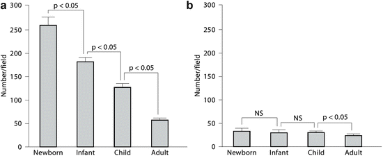
Fig. 11.3
Cell density in the human vocal fold mucosa. NS Not significant. (a) Vocal fold stellate cell density in the human macula flavae. (b) Fibroblast density in the lamina propria (Reproduced from Sato et al. [12])
The appearance of collagenous and elastic fibers composing vocal ligament, in and between the macula flavae of adult human vocal folds, suggests the implication that the macula flavae control the synthesis of these fibers [19]. Mainly based on morphological investigations into the macula flavae, the role of vocal fold stellate cells in collagen and elastic fiber synthesis have hypothesized by several authors [17, 21, 22]. However, it is difficult to prove directly that vocal fold stellate cells synthesize fibrous proteins composing the vocal ligament, because the vocal ligament is unique to human vocal folds and the verification of the hypothesis is difficult. So far, there have been only histological observation and there have been no evidences based on prospective, controlled, or experimental study. The functions of macula flavae and stellate cells in human vocal fold development and maintenance are still to be elucidated.
11.4 What Stimulate Vocal Fold Development?
Among mammals, only humans can speak and only human adult vocal folds have layered structure such as vocal ligament and Reinke’s space [28]. What are the factors of initiating the development from pediatric vocal folds into adult vocal folds exhibiting specialized microanatomy is still in question.
Tension has been thought to be the most important factor that influences the synthesis of collagenous fibers by fibroblasts [29]. The bending stresses on the vocal folds associated with phonation are greatest in the region of the macula flavae [30]. Sato et al. examined human vocal folds that remained unphonated after birth [31–33]. They reported that the vocal folds do not have a vocal ligament, Reinke’s space, or a layered structure, and the stellate cells in macula flavae appeared to have decreased activity to produce extracellular matrix. Therefore, they suggested that the tensions caused by phonation after birth may stimulate the vocal fold stellate cells in the anterior and posterior macula flavae to accelerate production of extracellular matrix and form the vocal ligament, Reinke’s space, and layered structure [31–33].
However, several studies have shown that collagen and elastin fibers are observed in lamina propria of fetal vocal folds [15, 17, 19, 20]. Besides the influence of phonation, the genetic control of the fibrous protein syntheses is thought to be of considerable importance.
Vocal folds are known to be androgen and estrogen sensitive. These gonadal steroids influence juvenile vocal fold maturation and result in voice changes from childhood to adulthood that vary in females and males, particularly during puberty [34–36


Stay updated, free articles. Join our Telegram channel
Full access? Get Clinical Tree
