Velopharyngeal Insufficiency
Velopharyngeal insufficiency (VPI) refers to the abnormal sound resulting from the inability to separate the nasopharynx from the oropharynx during speech. It may be caused by a structural abnormality or neuromuscular condition. Both structural and neuromuscular causes may be congenital or acquired.
17-1 Pharyngeal Flap (Palatopharyngoplasty)
Introduction
Pharyngeal flap is an operation designed to correct velopharyngeal insufficiency (VPI) thereby creating a new structure that enables the patient to develop normal speech and resonance.
Indications
A pharyngeal flap is indicated when VPI significantly affects speech intelligibility and nasopharyngoscopy illustrates a consistent central defect in velopharyngeal sphincter closure.
Preoperative Evaluation
Optimally, a thorough assessment of a child with suspected VPI involves the collaborative efforts of a speech-language pathologist, an otolaryngologist, and a geneticist. Geneticists offer expertise in the identification of syndromes associated with VPI and frequently conduct DNA testing. They also screen for developmental delays, special educational needs, general pediatric problems, and problems with social development. The perceptual evaluation of speech remains the gold standard for determining the need for intervention. Nasopharyngoscopy is a minimally invasive endoscopic procedure that allows direct assessment of the velopharyngeal mechanism during speech; it is therefore performed with the patient awake and not sedated. Because the pharyngeal flap creates a partial obstruction of the upper airway, care must be taken to identify patients with other sources of airway obstruction so as not to cause postoperative obstructive sleep apnea (OSA).
Operative Technique
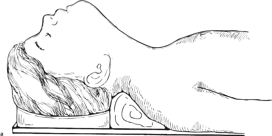
1. The pharyngeal flap is performed under general endotracheal anesthesia, utilizing an oral RAE tube. The patient is placed in the Rose position (Fig. 17.1a).
2. Palpation and visual inspection of the posterior pharyngeal wall are undertaken, looking for medialized carotid arteries.
3. The incisions are planned on the posterior pharyngeal wall. The lateral limbs of the incision are at the level of the posterior tonsillar pillars. If a wide flap is needed, the tonsillar pillars can be reflected laterally and the incisions can be placed in a more lateral position. The inferior aspect of the flap is generally at the level of the mid-tonsil (Fig. 17.1b).
4. The soft palate incision is made on the nasopharyngeal surface of the soft palate, ideally at the level of the velopharyngeal closure, which is generally 3 to 5 mm cephalad from the soft palate margin (Fig. 17.1c).
5. Proposed pharyngeal incision lines and the posterior aspect of the soft palate are infiltrated with 1% lidocaine with 1:100,000 units of epinephrine. Distortion of the tissue should be avoided.
6. The lateral incisions are carried down to the prevertebral fascia bilaterally. The inferior transverse incision is then made. The flap is elevated in this relatively avascular plane. The superior limit of elevation of the flap is high in the nasopharynx, at the level of velopharyngeal closure. This level is generally at the inferior edge of the adenoid remnant (Fig. 17.1d,e).
7. Closure of the donor site is undertaken. The inferior two-thirds of the donor site is closed with 3.0 Vicryl sutures (Ethicon). Extending the closure too far superiorly creates a stenosis of the nasal/oropharyngeal inlet. To minimize dead space beneath the repair, the superior suture should incorporate the prevertebral fascia when closing the mucosa (Fig. 17.1f).
8. A pocket is created on the posterior aspect of the soft palate (Fig. 17.1g,h). The incision must be at the level of velopharyngeal closure. The central aspect of the incision is made with a right angle Beaver blade. The palate is everted anteriorly with forceps. The incision is made into the substance of the palate. Care must be taken not to extend the incision inferiorly along the tonsillar pillars. The lateral aspect of the incision is made with palate scissors, again everting the palate for adequate visualization. One tine of the scissors is placed within the previously created pocket in the midline, and the tine is slid laterally along the plane of velopharyngeal closure. The scissors is turned such that the blades are perpendicular to the plane of the soft palate.
9. Endotracheal tube stents are placed transnasally into the hypopharynx; 3.5 endotracheal tubes are used for children age 6 years and younger and 4.0 tubes are used for those 8 years and older. For children from 6 to 8 years of age, endotracheal tube size is determined by the size and weight of the child. The stents define the size of the lateral ports (Fig. 17.1h,i,j).
10. Starting with the lateral sutures, the pharyngeal flap is positioned into the soft palate. A 3.0 Vicryl suture is placed laterally, going through the oral surface of the soft palate and exiting the previously created fishmouth incision in the soft palate (Fig. 17.1j).
11. The suture is placed in the pharyngeal flap at the junction of the proximal and middle thirds of the flap. The suture is placed in the muscle and submucosa only; the squamous mucosa is not included in this suture.
12. The suture then passes through the fishmouth incision of the soft palate and exits into the oral cavity within 2 mm of its initial pass, completing the horizontal mattress suture (Fig. 17.1k).
13. The suture is pulled (not tied) to position the flap into the palate. A mirror is used to check the size of the lateral port created when the flap is inset into the soft palate. The flap should touch the endotracheal tube, leaving space anterior and posterior to the endotracheal tube stent. The flap should not wrap tightly around the stent, as nasopharyngeal obstruction will likely develop. The suture is then loosened, and the opposite lateral suture is placed in similar fashion and checked (Fig. 17.1l).
14. Two paramedian sutures are similarly placed to finish the insetting of the flap.
15. The sutures are tied, starting with the lateral sutures. Care must be taken to approximate the flap into the pocket without strangulating the tissue. Care also must be taken to ensure that the free end of the flap is positioned such that the muscle of the flap is in contact with the raw surface of the soft palate. The squamous mucosa of the flap cannot reside within the soft palate pocket.
16. A final check of the position of the flap and size of the ports is undertaken. The flap should not be visible from the oral cavity, as it needs to be high in the nasopharynx.
17. The endotracheal tube stents are positioned in the oropharynx, with the tips at the level of the mid-tonsil (Fig. 17.1l).
18. The endotracheal tubes are secured to the upper lip with tape and then trimmed.
19. A number 6-8 Fr suction catheter is measured to direct the suctioning of the stents postoperatively. The catheter should extend 5 mm beyond the tip of the stent to ensure continued patency of the tube.
20. Elbow cuffs are used to protect the nasal stents.
21. Stents are removed the following morning. The child is discharged after demonstrating an adequate airway and adequate oral intake; this usually occurs 48 hours postoperatively.
22. Steroids should be avoided, as they may increase the risk of dehiscence of the flap from the soft palate pocket.
23. Speech therapy should begin 3 weeks postoperatively to obtain maximal functional use of the pharyngeal flap.
Complications
1. Flap breakdown
2. Persistent VPI
3. Inadequate nasal airway