(1)
Department of Pathology, Duke University Medical Center, Durham, NC, USA
Abstract
The uveal tract comprises the iris, the ciliary body, and the choroid, each of which has distinct histological features.
A significant component of uveal tract pathology derives from its population of indigenous melanocytes. The majority of the eyes enucleated for a uveal lesion that you are likely to see on the surgical pathology bench are malignant melanoma.
This chapter will focus on uveal melanoma and two other less common tumors: ciliary body medulloepithelioma and melanocytoma (also known as the magnocellular nevus).
A few other lesions that enter the differential diagnosis will also be introduced.
The uveal tract comprises the iris, the ciliary body, and the choroid, each of which has distinct histological features.
A significant component of uveal tract pathology derives from its population of indigenous melanocytes.
The majority of the eyes enucleated for a uveal lesion that you are likely to see on the surgical pathology bench are malignant melanoma.
This chapter will focus on uveal melanoma and two other less common tumors: ciliary body medulloepithelioma and melanocytoma (also known as the magnocellular nevus).
A few other lesions that enter the differential diagnosis will also be introduced.
Iris
The iris is the tissue that gives color to the eye. A wide spectrum of eye color is possible, and the color of one’s eye is determined by a complex interaction of genetics and the number and size of melanin pigment granules in the iris stromal melanocytes.
The anterior border of the normal iris has an undulating or crypt-like appearance (Fig. 5.1).
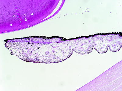
Fig. 5.1
The normal iris has an anterior crypt-like and undulating appearance. The iris pigment epithelium lines the posterior surface of the iris. The lens is in the top left of the figure, and the cornea stroma is seen in the bottom right
There are two smooth muscles in the iris that regulate the size of the pupil and therefore control the amount of light that enters the eye through the pupil. The pupil is a hole or an aperture (usually round) in the iris; it is not a histological tissue. The pupillary sphincter muscle borders the pupil (Fig. 5.2), and the thin dilator muscle lies immediately anterior to the double layer of iris-pigmented epithelium (Fig. 5.3).
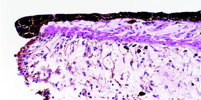
Fig. 5.2
Iris with the pupillary sphincter muscle at the edge of the iris leaflet
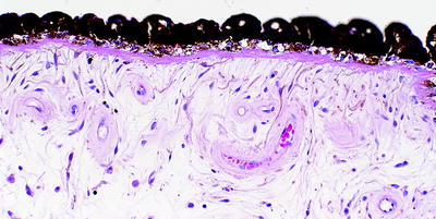
Fig. 5.3
Iris dilator muscle adjacent to the iris pigment epithelium
The normal blood vessels within the iris stroma have a cuff of collagen fibers that give them a thickened appearance; do not mistake this as hypertensive vascular disease (Fig. 5.4).
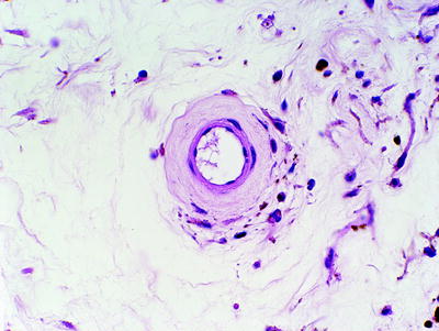
Fig. 5.4
Normal iris blood vessel with a collagenous cuff
Some histological abnormalities of the iris include:
Ectropion uveae: when the iris pigmented epithelium extends along the anterior surface of the iris. This may be associated with neovascular glaucoma.
Descemetization: when Descemet’s membrane extends along the anterior surface of the iris.
Ciliary Body
The ciliary body is responsible for the production of aqueous humor.
The ciliary body components include:
Smooth muscle (Fig. 5.5), which has a triangular shape in the horizontal plane (see Fig. 1.3). The smooth muscle of the ciliary body assists with accommodation of the lens by relaxing or contracting to change the shape of the lens.
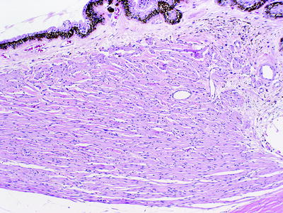
Fig. 5.5
Ciliary body with its smooth muscle component. The junction of the pars plicata and the flattened pars plana is seen at the top left of the image
Pars plicata with ciliary processes and an inner nonpigmented epithelial layer and an outer pigmented epithelial layer (Fig. 5.6).
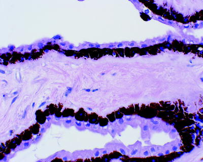
Fig. 5.6
Ciliary body pars plicata processes with an inner nonpigmented epithelial layer and an outer pigmented epithelial layer (inner refers to being closer to the vitreous cavity)
Pars plana, the flattened component of the ciliary body and the outer pigmented layer of which joins the retinal pigment epithelium.
Choroid
The choroid extends from the ora serrata (the junction of the pars plana of the ciliary body and the anterior border of the retina) to the optic nerve head and is located between the retina and the sclera.
The choroid is a pigmented and vascularized tissue, and its dark color was thought to resemble a grape (uva), thus the terminology uveal tract.
Within the choroid are melanocytes, a capillary network known as the choriocapillaris, and Bruch’s membrane (Figs. 5.7 and 5.8).
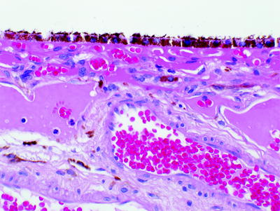
Fig. 5.7
Choroid with melanocytes and vessels. The retinal pigment epithelium is at the top of the image
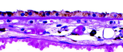
Fig. 5.8
Choroid with Bruch’s membrane immediately beneath the retinal pigment epithelium
Ganglion cells are occasionally seen within the choroid (Fig. 5.9).
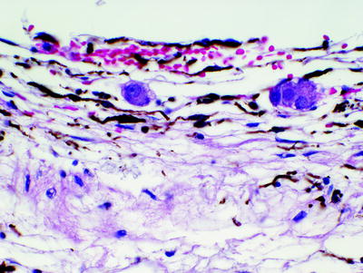
Fig. 5.9
A few ganglion cells are present in the normal choroid
Uveal Melanoma
Intraocular malignant melanomas arise from melanocytes of the uveal tract (iris, ciliary body, and choroid).
Uveal melanoma is the most common primary intraocular malignant neoplasm in the USA and is most commonly seen in adult Caucasian males.
Uveal melanomas can present clinically with visual complaints, can be detected incidentally on ocular examination, and sometimes are evident by simply looking at the eye (Figs. 5.10, 5.11).
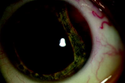
Fig. 5.10
Clinical image showing a ciliary body melanoma compressing the iris
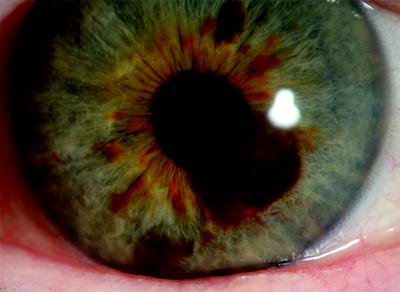
Fig. 5.11
Clinical image showing a melanoma arising from the iris
B-scan ultrasound (Fig. 5.12) and CT scanning (Fig. 5.13) or MR imaging can be used to assist with the preoperative diagnosis.
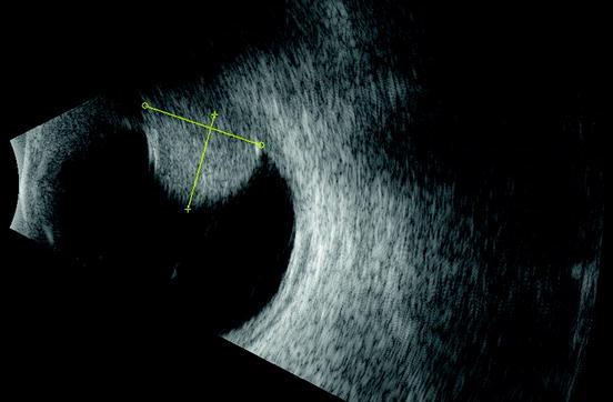
Fig. 5.12
B-scan ultrasound showing a choroidal melanoma
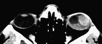
Fig. 5.13
Computerized tomography (CT) scan showing a ciliochoroidal melanoma with associated retinal detachment
Uveal melanomas are much more commonly encountered in the choroid, followed by the ciliary body, and are least frequent in the iris.
There is a relationship with nevi, congenital melanosis oculi, and oculodermal melanocytosis (nevus of Ota).
Bilateral diffuse uveal melanocytic proliferation (BDUMP) is a paraneoplastic syndrome associated with primary carcinomas of the lung, pancreas, and other internal organs.
Melanomalytic glaucoma is when uveal melanomas block the anterior chamber angle and extend into Schlemm’s canal (Figs. 5.14, 5.15, 5.16). Melanoma will rarely line the posterior surface of Descemet’s membrane (Fig. 5.17).
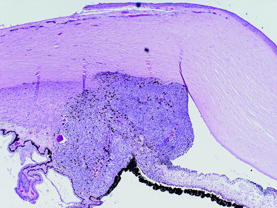
Fig. 5.14
Ciliary body melanoma blocking the anterior chamber angle and infiltrating the iris
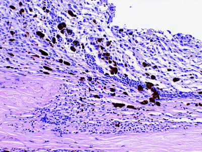
Fig. 5.15
Ciliary body melanoma extending into Schlemm’s canal
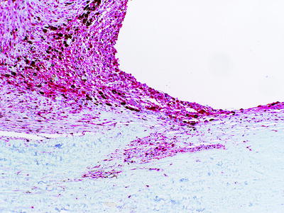
Fig. 5.16
Ciliary body melanoma extending into Schlemm’s canal. The tumor cells are highlighted with a Mart-1 immunohistochemical stain
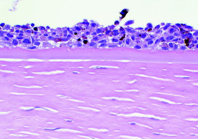
Fig. 5.17
Malignant melanoma lining the posterior surface of Descemet’s membrane
Neovascularization of the iris is characterized by small capillaries along the anterior border of the iris when neovascular glaucoma is present.
Uveal melanoma may extend through the sclera via emissary channels (Fig. 5.18) and involve extraocular muscles (Fig. 5.19).
< div class='tao-gold-member'>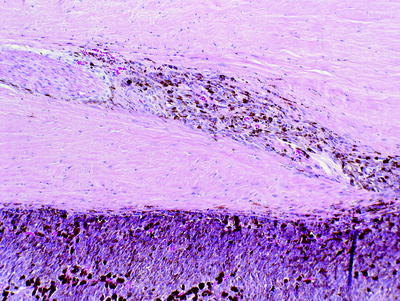 Only gold members can continue reading. Log In or Register to continue
Stay updated, free articles. Join our Telegram channel

Full access? Get Clinical Tree


