Purpose
To report ultrasound biomicroscopic (UBM) findings of iris-sutured foldable posterior chamber intraocular lenses (PCIOLs).
Design
Prospective, noninterventional consecutive case series.
Methods
Fifteen eyes with foldable acrylic IOL implantation using peripheral iris suture fixation in the absence of capsular support were included. UBM was used to determinate the haptic position in relation to the ciliary sulcus and ciliary body in these eyes. Additionally, anterior chamber depth, lens tilt, site of suture fixation, focal iris or angle abnormalities, and relationship of iris to lens were determined. Main outcome measures were haptic position, anterior chamber depth, and iris anatomic changes.
Results
Of the 30 haptics imaged, 16 (53.3%) were positioned in the ciliary sulcus. Nine (30%) haptics were found over the ciliary processes, and 5 (16.7%) were over pars plana. No patients were found to have peripheral anterior synechiae present at the haptic position. The mean (± standard deviation) depth of the anterior chamber was 3.84 ± 0.36 mm. The iris profile was altered in all patients at the iris–haptic suture fixation site. No angle abnormalities or tilted lenses were found.
Conclusions
Iris-sutured PCIOL haptics were found to be in the ciliary sulcus or over the ciliary body with no significant tilt on UBM analysis. The procedure respects the angle anatomy, and no evidence of angle closure was found. The anterior chamber was deeper than has been reported previously for scleral sutured PCIOLs and was similar to that of pseudophakic eyes. This may have implications for surgical technique, IOL power calculations, and postoperative complications.
Intraocular approaches to correct aphakia in cases of inadequate capsular support include an anterior chamber intraocular lens (ACIOL), a transscleral fixated posterior chamber intraocular lens (PCIOL), or an iris-sutured PCIOL. An American Academy of Ophthalmology device review concluded that all 3 options are comparably safe and effective. However, precise determination of small differences in visual outcome or complication rates requires a large prospective, randomized clinical trial.
Despite improvements in design, ACIOLs historically have been associated with angle complications, including corneal decompensation, glaucoma, and uveitis. Scleral-sutured IOLs, although reducing some of the concerns of ACIOLs, have been associated with IOL tilt, suture breakage, and endophthalmitis.
Iris-sutured IOLs have comparative advantages over the other options, including reduced inflammation, absence of suture exposure risk, and respect for angle structures. With adaptation to foldable IOLs, this technique has been reported with recent enthusiasm. However, one of the criticisms of this IOL fixation position is concern for close uveal contact and potential associated complications.
The ultrasound biomicroscope operates at a frequency of 50 MHz, producing images with a resolution of approximately 40 μm. This provides a unique ability to assess anatomic relationships between structures in the anterior segment of the eye and thus is ideally suited to study iris-sutured PCIOLs. Specifically, the ultrasound biomicroscope has the ability to assess haptic position in relation to the sulcus and ciliary body, anterior chamber depth (ACD), vitreous incarceration, focal iris abnormalities, angle anatomy, and relationship of the iris to lens (straight or tilted). The purpose of this study was to present ultrasound biomicroscopy (UBM) findings of iris-fixated foldable IOLs implanted for aphakia.
Methods
A prospective UBM evaluation of 15 eyes that underwent iris suture fixation of an acrylic foldable IOL for management of aphakia was performed. Surgery was performed by 1 of 3 surgeons (I.I.K.A., C.F.K., G.P.C.), with the technique described by Condon and by Stutzman and Stark, which is summarized briefly below. No intraocular complications occurred in any of the eyes enrolled.
Under topical anesthesia a 3-piece acrylic PCIOL (AcrySof MA60AC; Alcon Laboratories, Fort Worth, Texas, USA) was folded and inserted through a 3.5-mm clear corneal tunnel incision. Once in the anterior chamber (AC), both haptics were projected through the pupil while the optic was held just above the iris plane. The IOL was unfolded slowly, allowing the haptics to extend behind the posterior iris surface while the optic, supported by the spatula above the iris plane, was captured completely by the pupil and stabilized.
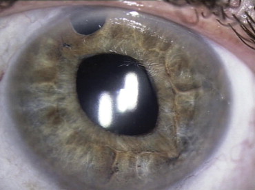
To fixate each haptic to the peripheral iris, a 10–0 polypropylene suture on a long curved CIF-4 needle (Ethicon, Inc, Somerville, New Jersey, USA) or PC-7 needle (Alcon Laboratories) was passed through peripheral iris, behind the haptic, and out through the iris and peripheral cornea. The Siepser slipknot technique or microtying forceps (Ahmed Microtying Forceps; MicroSurgical Technology, Redmond, Washington, USA) were used to tie the suture. The second haptic was secured in the same manner. The optic then was prolapsed into the posterior chamber ( Figure 1 ). When it was required, anterior or posterior vitrectomy, or both, was performed to clear the retropupillary space before IOL implantation.
Two physicians (C.J.P., H.I.), using a commercial version of the ultrasound biomicroscope (Humphrey Instruments; San Leandro, California, USA), conducted all UBM examinations. UBM imaging procedures have been described elsewhere.
The central ACD, iris profile, iris-haptic fixation sites, AC angle, IOL optic tilt and position, and haptic position were examined with the UBM in all eyes. Haptic position was designated as CS if located in the ciliary sulcus, CP if located over the ciliary processes, and PP for haptics located over the pars plana.
Results
The mean age of enrolled patients ranged from 20 to 86 years; mean (± standard deviation) was 51.8 ± 21.8 years. There were 4 eyes from females and 11 from males. Follow-up duration ranged from 3 to 33 months; the mean (± standard deviation) follow-up was 10.5 ± 9.7 months.
Fourteen eyes improved their best-corrected visual acuity (BCVA), whereas 1 eye maintained BCVA. The mean preoperative BCVA was 0.72 ± 0.47 logarithm of the minimal angle of resolution units, and the postoperative BCVA was 0.38 ± 0.19 logarithm of the minimal angle of resolution units ( P = .001).
UBM analysis was performed on 15 eyes. UBM examination found that 16 haptics (53.3%) were located in the ciliary sulcus ( Figure 2 ), 9 haptics (30%) were over the ciliary processes ( Figure 3 ), and 5 haptics (16.7%) were over pars plana ( Figure 4 ). No haptics were found anterior to the sulcus region. Figure 5 summarizes the actual position of all haptics imaged. The location and relative position of haptics (clock hours) are detailed in Table 1 . We did not find any alteration in angle anatomic features, nor any peripheral anterior synechiae nor any degree of angle closure. The mean ACD was 3.84 ± 0.36 mm (range, 3.17 to 4.5 mm).
| Patient No. | ACD | Haptic 1 | Haptic 2 | Site of Suture | ||
|---|---|---|---|---|---|---|
| 1 | 4.05 | CS | 1:00 | CP | 7:00 | Mid periphery |
| 2 | 3.17 | CP | 6:00 | CP | 12:00 | Mid periphery |
| 3 | 3.61 | CS | 1:00 | CS | 7:00 | Mid periphery |
| 4 | 3.40 | CS | 12:00 | CP | 6:00 | Mid periphery |
| 5 | 4.50 | CP | 6:00 | PP | 12:00 | Mid periphery |
| 6 | 3.59 | CS | 6:00 | CS | 12:00 | Mid periphery |
| 7 | 3.89 | CS | 3:00 | CS | 9:00 | Mid periphery |
| 8 | 3.75 | CS | 10:00 | PP | 4:00 | Mid periphery |
| 9 | 3.70 | CS | 11:00 | PP | 5:00 | Mid periphery |
| 10 | 3.96 | CS | 3:00 | CS | 9:00 | Mid periphery |
| 11 | 4.33 | CP | 11:00 | CP | 7:00 | Mid periphery |
| 12 | 3.67 | CS | 12:00 | CP | 6:00 | Mid periphery |
| 13 | 4.20 | PP | 3:00 | PP | 9:00 | Mid periphery |
| 14 | 3.63 | CS | 2:00 | CS | 7:00 | Mid periphery |
| 15 | 4.22 | CS | 12:00 | CP | 6:00 | Mid periphery |
All the IOLs were in a planar position; no IOL tilt was found in any patient. No synechia or vitreous incarceration was evident during UBM examination. At the point of iris–haptic suture fixation, there was a focal acute alteration in the iris profile ( Figure 6 ). Other than the point of iris–haptic fixation, there was no further contact of the IOL optic or haptic to the posterior iris.
Results
The mean age of enrolled patients ranged from 20 to 86 years; mean (± standard deviation) was 51.8 ± 21.8 years. There were 4 eyes from females and 11 from males. Follow-up duration ranged from 3 to 33 months; the mean (± standard deviation) follow-up was 10.5 ± 9.7 months.
Fourteen eyes improved their best-corrected visual acuity (BCVA), whereas 1 eye maintained BCVA. The mean preoperative BCVA was 0.72 ± 0.47 logarithm of the minimal angle of resolution units, and the postoperative BCVA was 0.38 ± 0.19 logarithm of the minimal angle of resolution units ( P = .001).
UBM analysis was performed on 15 eyes. UBM examination found that 16 haptics (53.3%) were located in the ciliary sulcus ( Figure 2 ), 9 haptics (30%) were over the ciliary processes ( Figure 3 ), and 5 haptics (16.7%) were over pars plana ( Figure 4 ). No haptics were found anterior to the sulcus region. Figure 5 summarizes the actual position of all haptics imaged. The location and relative position of haptics (clock hours) are detailed in Table 1 . We did not find any alteration in angle anatomic features, nor any peripheral anterior synechiae nor any degree of angle closure. The mean ACD was 3.84 ± 0.36 mm (range, 3.17 to 4.5 mm).


