Purpose
To report the results of a 2-year follow-up study of Japanese polypoidal choroidal vasculopathy (PCV) patients treated with reduced-fluence photodynamic therapy (PDT) monotherapy.
Design
Prospective interventional case series.
Methods
In the present study, 38 eyes of 38 consecutive patients underwent PDT with a reduced laser fluence of 25 J/cm 2 . During the 2-year follow-up, visual acuity (VA) and optical coherence tomography measurements were performed every 3 months after the PDT procedure and then compared with baseline values. PCV vascular lesions were evaluated by indocyanine green and fluorescein angiography.
Results
At baseline, the mean logarithm of the minimal angle of resolution (logMAR) best-corrected VA (BCVA) was 0.43. There was a significant improvement of the mean logMAR BCVA to 0.28 and 0.29 at 12 and 24 months, respectively ( P < .0001, P = .001). The logMAR BCVA was stable or improved by ≥0.3 in 36 (95%) of the eyes at the 2-year follow-up. In 13 eyes in which the baseline VA was better than 20/40, there was a significant improvement of the mean logMAR BCVA at 12 months, with the acuities continuing to be stable at 24 months. The mean number of treatment sessions during the 24-month study period was 1.9.
Conclusions
Reduced-fluence PDT monotherapy for PCV effectively improved and maintained the VA over a 24-month period, even in eyes with a baseline VA better than 20/40. In addition, the number of treatments could be much smaller as compared with intravitreal injection of anti–vascular endothelial growth factor agents.
When data from the Japanese Age-Related Macular Degeneration Trial (JAT) were compared with data from the Treatment of Age-Related Macular Degeneration with Photodynamic Therapy (TAP) study, a superior efficacy of the photodynamic therapy (PDT) monotherapy was seen in Japanese vs white subjects. Guidelines for the use of verteporfin in Japan demonstrated that after administration of this therapy, the visual acuity (VA) was effectively maintained in all lesion types for at least 12 months. This previous study also showed there was a significant improvement in the VA of eyes with polypoidal choroidal vasculopathy (PCV) ( P < .001), the prevalence of which is postulated to be higher in Asian populations than in white. However, there was a decline in the VA in eyes that had a baseline VA that was better than 20/40.
Several studies have shown that PDT damages the physiological choriocapillary layer beyond the irradiated area, with repeated PDT leading to persistent choriocapillary nonperfusion in most eyes. Choroidal ischemia has been demonstrated to consistently induce a secondary angiogenic response along with an increased expression of vascular endothelial growth factor (VEGF). It is possible that verteporfin therapy may influence the physiological choroid and be the reason for further vision loss and the need for repeated treatments. The Visudyne in Minimally Classic Choroidal Neovascularization (VIM) study group demonstrated that the proportion of patients having an acute severe vision decrease did not differ between the reduced-fluence and the standard-fluence groups. Michels and associates reported that reduced-fluence PDT led to much less damage to the choriocapillaris as compared with standard-fluence treatment.
Since several previous studies have reported standard-fluence PDT was an efficacious treatment for PCV eyes, we studied the 1-year results of reduced-fluence PDT for PCV in Japanese patients. Our analyses showed that there was a significant improvement of vision in all of the PCV patient eyes, including those eyes that had a baseline VA better than 20/40. Recently, a few studies reported that, although the standard-fluence PDT for PCV improved or maintained the VA at 12 months, the mean VA decreased in conjunction with an increased duration of follow-up. Therefore, the current study was designed to further investigate the length of the efficacy related to this treatment.
Methods
A total of 38 eyes of 38 consecutive PCV patients (34 men, 4 women) who had undergone no previous treatment for PCV were enrolled in this prospective study between July 1, 2007 and June 30, 2009. All 38 patients were Japanese, with a median age of 71.0 years (range, 47–83 years). The diagnosis of PCV was made based on the fundus examination and indocyanine green angiography (ICGA) findings of elevated orange-red lesions, characteristic polypoidal lesions, and abnormal vascular networks. All patients received a verteporfin (Visudyne; Novartis Pharma, Tokyo, Japan) injection at a concentration of 6 mg/m 2 of body surface area over a 10-minute period. Five minutes after completion of the infusion, patients then underwent PDT with a light fluence of 25 J/cm 2 using Visulus PDT system 690S (Carl Zeiss Meditec AG, Jena, Germany). The laser spot size chosen covered all of the PCV vascular lesions, including the polypoidal lesions and the branching vascular network vessels on the ICGA, with an extra margin of 1000 μm added to ensure the coverage was complete. All patients were examined prior to the PDT (baseline) and then during regular follow-up visits every 3 months after the PDT. Best-corrected VA (BCVA) and optical coherence tomography (OCT) using an OCT3000 or Cirrus HD-OCT system (Carl Zeiss Meditec, Dublin, California, USA) were performed at each visit. Fluorescein angiography (FA) using the TRC50IX system with ImageNet2000 (Topcon, Tokyo, Japan) and ICGA using a Heidelberg Retina Angiograph 2 (HRA2) (Heidelberg Engineering GmbH, Heidelberg, Germany) were performed before the PDT, at 3 months after PDT, and at all time points where recurrence of exudative changes was determined by OCT. PCV vascular lesions were evaluated by ICGA, while FA was used to document leakage from polypoidal vascular lesions. The PDT endpoint was reached when FA showed a complete absence of leakage and OCT demonstrated an absence of exudative changes, including serous retinal detachment and retinal edema. When eyes showed no exudative changes on OCT but minor leakage on FA, these eyes were continued to be followed, but without any further treatment. Treatment safety was assessed based on the BCVA and adverse events. The BCVA was obtained using Landolt ring tests and was then converted into the logarithm of the minimal angle of resolution (logMAR). A significant BCVA improvement was defined as an increase of ≥0.3 logMAR units, while a worsening of the BCVA was defined as a decrease of ≤0.3 logMAR units. The BCVA outcome comparisons to baseline were performed using a paired t test.
Results
Patient characteristics are shown in the Table .
| Case No. | Age | VA | ICGA Findings at 3 Months | Number of Treatments | Subretinal Hemorrhage After PDT | |||
|---|---|---|---|---|---|---|---|---|
| At Baseline | 12 Months | 24 Months | Branching Vascular Network | Polypoidal Vascular Lesions | ||||
| 1 | 61 | 20/250 | 20/120 | 20/120 | Persistent | Disappeared | 1 | — |
| 2 | 77 | 20/30 | 20/20 | 20/30 | Disappeared | Disappeared | 3 | — |
| 3 | 82 | 20/60 | 20/25 | 20/40 | Persistent | Disappeared | 1 | 1 DA; disappeared at 3 mo |
| 4 | 47 | 20/20 | 20/10 | 20/10 | Disappeared | Disappeared | 1 | — |
| 5 | 59 | 20/30 | 20/20 | 20/13 | Disappeared | Disappeared | 1 | — |
| 6 | 59 | 20/200 | 20/100 | 20/100 | Persistent | Disappeared | 1 | — |
| 7 | 73 | 20/50 | 20/100 | 20/60 | Persistent | Existent | 3 | — |
| 8 | 77 | 20/50 | 20/22 | 20/60 | Persistent | Disappeared | 2 | 1 DA; disappeared at 6 mo |
| 9 | 75 | 20/40 | 20/60 | 20/50 | Persistent | Disappeared | 2 | — |
| 10 | 71 | 20/40 | 20/22 | 20/28 | Disappeared | Disappeared | 2 | — |
| 11 | 55 | 20/30 | 20/16 | 20/16 | Persistent | Disappeared | 1 | — |
| 12 | 77 | 20/30 | 20/25 | 20/60 | Persistent | Disappeared | 2 | — |
| 13 | 71 | 20/40 | 20/30 | 20/50 | Persistent | Disappeared | 4 | — |
| 14 | 64 | 20/28 | 20/30 | 20/25 | Persistent | Disappeared | 1 | — |
| 15 | 77 | 20/30 | 20/25 | 20/25 | Persistent | Disappeared | 1 | — |
| 16 | 68 | 20/28 | 20/16 | 20/20 | Persistent | Disappeared | 1 | — |
| 17 | 75 | 20/28 | 20/28 | 20/30 | Persistent | Disappeared | 1 | — |
| 18 | 79 | 20/120 | 20/120 | 20/100 | Persistent | Disappeared | 2 | — |
| 19 | 68 | 20/50 | 20/28 | 20/25 | Persistent | Disappeared | 1 | 0.25 DA; disappeared at 1 mo |
| 20 | 70 | 20/60 | 20/28 | 20/28 | Persistent | Disappeared | 1 | — |
| 21 | 83 | 20/60 | 20/28 | 20/20 | Persistent | Disappeared | 1 | — |
| 22 | 78 | 20/50 | 20/22 | 20/22 | Persistent | Disappeared | 1 | 0.5 DA; disappeared at 6 mo |
| 23 | 70 | 20/200 | 20/120 | 20/60 | Disappeared | Disappeared | 1 | 2.5 DA; disappeared at 3 mo |
| 24 | 71 | 20/100 | 20/50 | 20/50 | Persistent | Disappeared | 2 | — |
| 25 | 74 | 20/30 | 20/50 | 20/30 | Persistent | Disappeared | 4 | — |
| 26 | 80 | 20/100 | 20/120 | 20/500 | Persistent | Disappeared | 3 | — |
| 27 | 71 | 20/60 | 20/30 | 20/22 | Persistent | Disappeared | 3 | — |
| 28 | 71 | 20/60 | 20/100 | 20/60 | Persistent | Disappeared | 1 | — |
| 29 | 68 | 20/50 | 20/22 | 20/16 | Persistent | Disappeared | 1 | — |
| 30 | 73 | 20/50 | 20/60 | 20/40 | Persistent | Disappeared | 3 | — |
| 31 | 77 | 20/28 | 20/20 | 20/22 | Persistent | Disappeared | 1 | — |
| 32 | 65 | 20/40 | 20/16 | 20/16 | Persistent | Disappeared | 3 | — |
| 33 | 57 | 20/286 | 20/100 | 20/120 | Disappeared | Disappeared | 2 | — |
| 34 | 81 | 20/28 | 20/16 | 20/20 | Persistent | Disappeared | 1 | — |
| 35 | 74 | 20/50 | 20/16 | 20/25 | Persistent | Disappeared | 3 | — |
| 36 | 70 | 20/30 | 20/40 | 20/40 | Persistent | Disappeared | 4 | — |
| 37 | 78 | 20/30 | 20/25 | 20/28 | Persistent | Existent | 3 | — |
| 38 | 67 | 20/100 | 20/100 | 20/120 | Persistent | Existent | 3 | — |
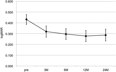
Visual Outcomes
The logMAR BCVA (mean ± standard deviation) was 0.43 ± 0.28 at baseline, 0.32 ± 0.32 at 3 months, 0.30 ± 0.31 at 6 months, 0.28 ± 0.32 at 12 months, and 0.29 ± 0.34 at 24 months. When compared with baseline values, significant changes in the BCVA were seen at 3, 6, 12, and 24 months ( P = .0014, P = .0006, P < .0001, and P = .001, respectively) ( Figure 1 ). In 36 of the 38 eyes (95%), the logMAR BCVA measurements at 24 months were improved by ≥0.3 or remained unchanged. Furthermore, in all 13 of the eyes that had a baseline VA that was better than 20/40, there was a significant improvement of the logMAR BCVA, from 0.18 ± 0.07 at baseline to 0.05 ± 0.20 at 12 months ( P = .015), with the values continuing to be stable until 24 months (logMAR BCVA: 0.09 ± 0.22).