The deviated nasal dorsum is a complex problem with a variety of proposed solutions. Straightening the deviated nose should be focused on maximizing cosmetic outcome while preserving or improving nasal function. Deviations can occur in one or a combination of the nasal thirds. A simple approach to treatment is to develop a strategy for each third of the nose. Tailoring maneuvers to alleviate problems in each specific third helps the surgeon deal with deviations in an effective and straightforward manner.
The deviated dorsum creates the appearance of a twisted, asymmetric, or deviated nose. This situation may create functional and aesthetic issues that require correction. Correction of the dorsal deviation may present itself as a challenge to the rhinoplasty surgeon. The dorsum is a complex three-dimensional structure in which correcting a functional issue may have an impact on cosmesis and cosmetic correction may impair nasal function. Therefore, any attempt at straightening a dorsum should strive to maximize cosmetic outcome and maintain or improve nasal function. This can be a difficult task for the surgeon.
A proper skeletal support is necessary to provide long-term aesthetic and functional results. In the past, rhinoplasty was typically a reductive operation commonly resulting in loss of support, whereas the modern operation is focused on restructuring the nose. To give the nose an appearance of being straight, the existing architecture is realigned or grafts are used to provide a symmetric and straight appearance. Most often, both strategies are used to achieve a straight and symmetric nose. Overall, autogenous cartilage is the material most commonly used to restructure the nose and provides material with which to strengthen, augment, camouflage, and reposition the deviated dorsum. Sources of autogenous cartilage are the nasal septum, auricular conchal cartilage, and costal cartilage.
Patient assessment
Deviations range from a subtle “C”-shaped deformity to a severe twisted nose deformity. Systematic facial analysis and evaluation of the dorsum are critical to correcting a deformity of this type. Such an analysis is complicated by the fact that most faces, on close examination, are vertically or horizontally asymmetric. As such, aligning the nose perfectly with one side or half of the face may not make it symmetric with respect to the other side. For example, the inferior third of the face may not lie in direct midline alignment with the upper third of the face. Hence, making a dorsum that bisects those two midline points results in a nose angled to one side. These imperfections and asymmetries should be pointed out to the patient before any surgery.
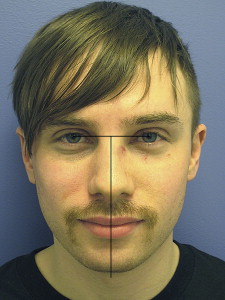
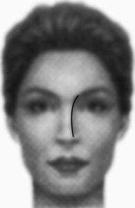
Perhaps the only consistent reference point for an frontal or anterior-posterior (A-P) photograph is the center point between the medial canthi on the nose with the head in the Frankfort plane. The other facial landmarks tend to be ineffective for analysis of dorsal symmetry because of facial asymmetries. A straight line is drawn from pupil to pupil. The center point between the medial canthi is marked. From this point, a straight midline vertical line is dropped intersecting the glabella, nasal dorsum, tip, columellar base, nasal spine, philtrum, upper incisors, and menton. Fig. 1 shows a patient with a very minimal upper third nasal deviation. In such cases where the deviation is subtle/minimal, a vertical line dropped from the centerpoint between the medial canthi or pupils can more clearly show this. Additionally, the rhinoplasty surgeon should analyze the patient’s brow-aesthetic line on the A-P photograph. The nasal width should provide a graceful curvilinear line from brow to nasal tip. Deviation of the dorsum alters the brow-tip aesthetic and affects the contour of this line. Fig. 2 shows an appropriate brow-tip aesthetic line for a patient with a straight nose.
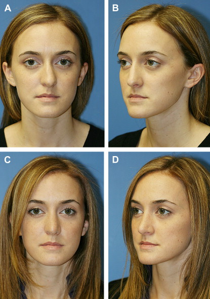
The upper, middle, and lower thirds of the dorsum are each analyzed independently and then together as a whole. The upper third is composed of the nasal bones and the ascending process of the maxilla. The middle third is composed of the dorsal septum fused in the midline to the paired upper lateral cartilages. The lower third is the nasal tip, which includes the lower lateral cartilages, caudal septum, and alar base. Each third is recorded as being deviated to the left or right. It is clinically significant whether the upper, middle, or lower third is deviated because the surgical management of each third of the vault has its own set of maneuvers to straighten it. In this article, the surgical approach to the deviated nose is based on this principle of thirds. Fig. 3 A , B shows a patient with dorsal deviation to the left. The upper third (nasal bones) is not deviated. The middle and lower thirds of the nose are deviated to the right, however. Fig. 3 C, D shows the patient after surgical straightening of the nose. These photographs were taken 6 months after surgery.
Integral to patient assessment is the assessment of the intended surgical approach. The approach for management of the deviated dorsum can be external or endonasal. The choice of approach used depends on the severity of the deviation and comfort level of the surgeon. The authors find that most maneuvers targeting the deviated nose can be comfortably performed with either approach. In the 1970s, Goodman and Charles introduced a columellar incision to provide unparalleled exposure and facilitate open rhinoplasty as a means to preserve anatomic structure. This dramatically decreased the learning curve of surgeons attempting to perform a complex procedure. Yet, this comes at the expense of potentially increased operative time, prolonged postoperative swelling, scar contracture, and loss of nasal tip support. There is also the issue of an external scar that may heal conspicuously depending on a variety of factors. The advantages of endonasal rhinoplasty include decreased operative time, rapid recovery, and less significant scar contracture. These benefits come at a cost of a more limited exposure, however.
Management of the upper third
Treatment of the deviated upper third is tailored to the upper third deformity. The nasal bones as a unit may be twisted to the side, but the ascending processes of the maxilla are uninvolved. There may be a fracture (rare) of the entire bony complex unit to one side. More commonly, there are variations on those extremes. The treatment is osteotomies, and the specific osteotomy is based on the identified abnormality.
Deviation of the bony pyramid is corrected with osteotomies made at specific locations along the vertical plane of the bones, depending on the location of bony deviation. Once the bony dorsum is mobilized, it may be manipulated and straightened, and occasionally stented. Osteotomies may be performed in a variety of ways. They can be performed using a continuous technique, which is the most common method used. In this type of osteotomy, the cuts are made in a continuous fashion, through the nasal sidewall. Osteotomies can also be performed in a perforating fashion, which is a postage stamp type of sequential fracture using a narrow osteotome (2–3 mm). Osteotomies can also be performed by means of percutaneous perforations, which are also performed with a narrow osteotome. The type of osteotomy is insignificant as long as there is a cut through the bone that allows sufficient bone movement for the bony pyramid to be shifted to midline. Lateral osteotomies are ideally performed in a high-low-high fashion. The initial part of the osteotomy begins at the pyriform aperture (high point) and travels laterally toward the ipsilateral eye. As the osteotomy nears the nasal process of the maxilla (low point), it travels cephalically and medially toward a point just medial to the medial canthus (high point).
Medial osteotomies are performed in addition to lateral (occasionally intermediate) osteotomies to allow increased mobility before manipulation of the bones. A combination of osteotomies is usually sufficient to bring the bones back to midline; however, lateral osteotomies alone may suffice. Medial osteotomies are performed by making osteotomies that begin just lateral to the septal insertion into the underside of the nasal bones. These osteotomies are aimed laterally to avoid rocker deformity.
Dorsal hump reduction may obviate the need for medial osteotomies. This is determined by how much hump is removed. When an open roof deformity is created after significant hump reduction, lateral osteotomies alone suffice because the central portion of the bony pyramid is absent. Because the bone is removed from the osteotomy, there is no central bone available to cut with an osteotome. If dorsal hump reduction does not remove a significant portion of the bony roof, greenstick fracture may result if lateral osteotomies alone are performed because there remains a bony bridge from the end of the lateral osteotomy to the root of the nose. Greenstick fractures are not ideal because they maintain a spring-like tendency, resulting in a return of the bony pyramid to its original configuration in the postoperative period.
Unilateral osteotomies can be performed in certain situations. The surgeon must be open to ways to reshape the upper third, including not only unilateral osteotomy but outfracture of nasal bones after osteotomy. The usual strategy of the facial plastic surgeon is to infracture nasal bones before trying to realign the nasal vault. In certain circumstances of severe deviation attributable to fracture, however, the pyramid is so deviated that a portion of bone must be outfractured.
In severe cases of deviation, a cross-root (or transverse-root) osteotomy can be performed. In this maneuver, a 2-mm osteotome can be used to make perforated percutaneous osteotomies in a horizontal fashion across the nasion, or just inferior to the nasal root. This series of perforations should connect the most cephalic aspect of the lateral osteotomies. This maneuver should provide tremendous mobility when combined with medial and lateral osteotomies. The bony dorsum can then be manipulated and molded with the opposite hand into place. This maneuver is usually used when medial and lateral osteotomies are insufficient in providing the mobility required for straightening.
Stay updated, free articles. Join our Telegram channel

Full access? Get Clinical Tree


