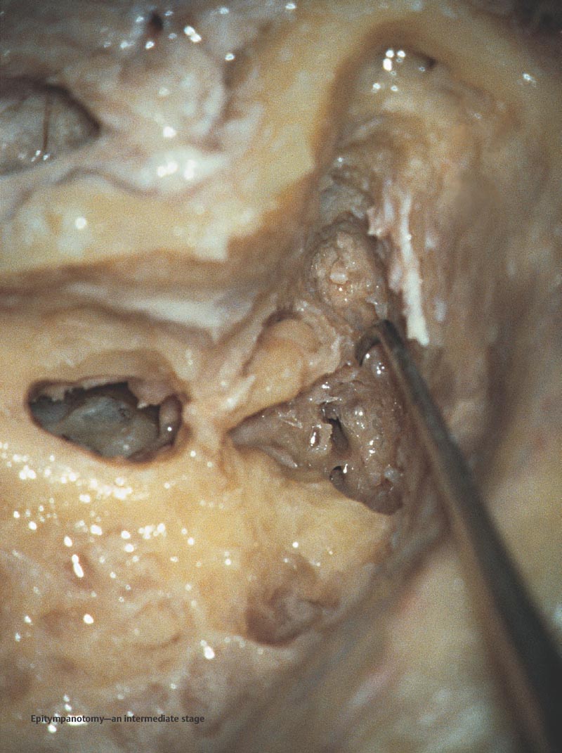
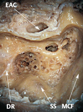
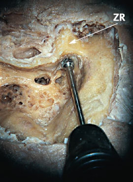
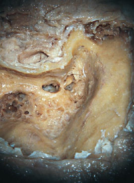
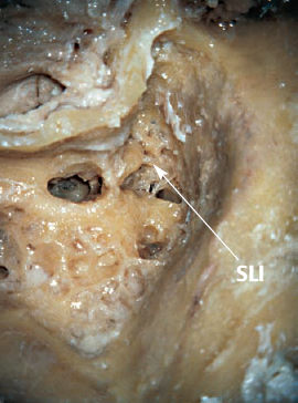
8 Transcortical Exposure of the Epitympanum The transcortical, full exposure of the epitympanum, generally carried out after a cortical mastoidectomy. Alternatively, it may be called epitympanotomy. The main indication is in the surgery for chronic otitis media with cholesteatoma. The cortical mastoidectomy and posterior tympanotomy have been completed. The temporal bone is ready for epitympanotomy. In chronic otitis media surgery, an epitympanotomy or epitympanectomy is carried out with or without a posterior tympanotomy, depending on the needs of the case. DR: Digastric ridge EAC: External auditory canal MCF: Middle cranial fossa (dural plate) SS: Sigmoid sinus (dural plate) The zygomatic root is identified and using a medium-sized cutting burr, the bone in this area is removed. Care should be taken not to damage the dura of the middle cranial fossa superiorly and the superior wall of the external auditory canal inferiorly. II: 3–4mm cutting burr MIL: Zygomatic root ZR: Zygomatic root Following extirpation of the lateral bone at the zygomatic root, the exposure is widened. The medial bone, which is close to the incus and malleus, is left untouched until the final stage of the procedure. Definitions and Tips
Definition
Indications
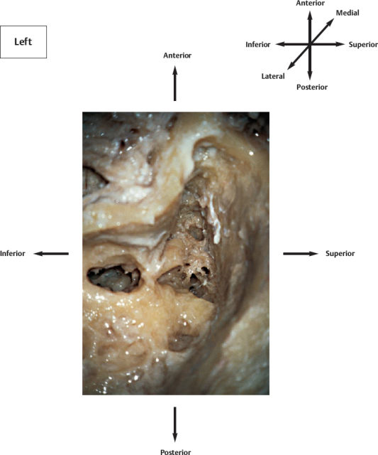
Anatomical Orientation
Surgical Steps
Transcortical Exposure of the Epitympanum
Only gold members can continue reading. Log In or Register to continue

Full access? Get Clinical Tree


