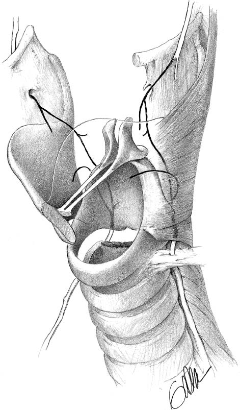
Thyroid and Parathyroid SurgeryGregory W. Randolph
The essential point to be made in a discussion of thyroid and parathyroid surgery is that meticulous surgical technique is paramount. This is crucial for appreciation of neck base anatomy, including recurrent laryngeal nerve and parathyroid recognition and preservation.
♦ Thyroid Surgery
Surgical Technique
Patient Positioning/Incision
- An inflatable thyroid bag or thyroid roll is placed under the shoulders, and important attention is given to adequate head support. Once the patient is positioned, the surgical bed is placed in a semi-sitting position to decrease venous pressure. The eyes are lubricated and taped.
- We typically employ 10 mg of Decadron® (dexamethasone) to help reduce the risk of neuropraxic nerve injury. With recurrent laryngeal nerve monitoring, muscle relaxation is avoided.
- A 4 to 5 cm collar-type thyroid incision is made one thumb breadth below the cricoid. An incision that is too low, especially if the neck is thin, can cause excessive scarring in the suprasternal notch region.
Subplatysmal Skin Flap
- A subplatysmal skin flap is raised to approximately the level of the superior thyroid cartilage. The anterior jugular veins are let down. It is infrequent that an inferior flap needs to be raised.
Identification of the Airway
- The strap muscles are dissected in the midline with identification of the medial edge of the sternohyoid muscles and sternothyroid muscles.
- The cricoid cartilage, the cricothyroid membrane, the thyroid cartilage, and the trachea above and below the isthmus are dissected. This allows identification of any prelaryngeal or pretracheal adenopathy and also allows identification of the pyramidal lobe should it be present.
Strap Muscles
- The strap muscles are reflected off the ventral surface of the thyroid gland. This is facilitated by medial retraction of the thyroid gland opposing the lateral retraction of the strap muscles. Strap muscles can be divided in cases of goiter.
- The middle thyroid vein is visualized, clamped, and tied with 2–0 silk.
Inferior Pole
- With slight upward and medial thyroid retraction and lateral strap muscle retraction, the inferior pole of the thyroid is dissected on a capsular plane. Usually several small inferior ventral thyroid veins can be clamped and tied.
- The inferior parathyroid typically resides in fat adjacent to the inferior pole, often in the thyrothymic horn itself. This fat and the inferior thyroid can be brushed away from the thyroid as these inferior thyroid pole veins are taken. A parathyroid gland that has good color, has not been dissected from the surrounding fat, and has a laterally oriented pedicle at the end of the case can be relied on for postoperative function (Fig. 18–1).
Figure 18–1 The inferior parathyroid can be found adjacent to the inferior pole in thyrothymic horn fat. (From Randolph G. Surgery of the Thyroid and Parathyroid Glands. Philadelphia: Saunders; 2003. Reprinted with permission.)
Recurrent Laryngeal Nerve
- This is best identified at the midpolar level. Recurrent laryngeal nerve stimulation can be performed through the overlying fascia to identify the nerve even prior to visualization. Gentle dissection with a snap parallel to the anticipated direction of the nerve is best.
Figure 18–2 The recurrent laryngeal nerve is shown in the tracheoesophageal groove, piercing the ligament of Berry and extending under the inferiormost fibers of the inferior constrictor. The left thyroid cartilage is removed in this illustration to show intralaryngeal nerve anatomy. (From Randolph G. Surgery of the Thyroid and Parathyroid Glands. Philadelphia: Saunders; 2003. Reprinted with permission.)
- The nerve in this area can be identified as it crosses above or below the inferior thyroid artery. The inferior thyroid artery in the lateral thyroid region can be identified after the middle thyroid vein has been taken through a gentle pulsation.
- The recurrent laryngeal nerve above the inferior thyroid artery may branch. All branches must be preserved. The recurrent laryngeal nerve does not need to be dissected along its entire path in this area. The dissection can be through several windows through which the path of the nerve is fully seen. This helps to preserve medially running branches of the inferior thyroid artery. The nerve at this point is dissected up to where it dives underneath the thyroid lobe, toward the ligament of Berry (Fig. 18–2).
Superior Parathyroid
- Before the superior pole is taken, the superior parathyroid is identified on the posterolateral aspect of the superior pole under fascia in this region. It generally rests in fat in this location. It is best to create a plane between the superior parathyroid posteriorly (i.e., dorsal) and the posterolateral aspect of the superior pole, which, when retracted, is ventral. If possible, we prefer to dissect the superior parathyroid away from the superior pole prior to taking superior pole vessels because we feel this may help preserve superior pole posterior branches, which may in part vascularize the superior parathyroid (Fig. 18–3).
Figure 18–3 The superior parathyroid is identified in fat adjacent to the lateral aspect of the superior pole directly lateral and dorsal to the recurrent laryngeal nerve entry site. (From Randolph G. Surgery of the Thyroid and Parathyroid Glands. Philadelphia: Saunders; 2003. Reprinted with permission.)
Superior Pole

Stay updated, free articles. Join our Telegram channel

Full access? Get Clinical Tree

