As more patients with Down syndrome are living into adulthood, attention has focused on health factors that affect the quality of the patient’s life and their ability to reach full potential. Patients with Down syndrome have several morphologic abnormalities that predispose them to problems with the ear, nose, and throat, and appropriate treatment can have a significant impact on the quality of life of these patients. Otolaryngologists are likely to see many patients with Down syndrome throughout their careers. This article reviews the literature to provide information and recommendations regarding management of Down syndrome.
- •
The child with Down syndrome should undergo behavioral audiologic testing every 6 months, or every 3 months if the patient has very stenotic ear canals. Treatment of hearing loss caused by recurrent otitis media and otitis media with effusion (OME) should be aggressive, with close follow-up. Parents should be prepared for multiple sets of pressure equalization (PE) tubes throughout the child’s life.
- •
Up to 80% of patients with Down syndrome have obstructive sleep apnea. All children with Down syndrome should get an overnight polysomnography study between the ages of 3 and 4 years. Parents are not reliable in assessing sleep apnea.
- •
Primary treatment of obstructive sleep apnea syndrome (OSAS) is tonsillectomy and adenoidectomy, but parents should be prepared that this is curative in only 25% to 50% of children with Down syndrome, and that their child may require further surgery to alleviate obstruction, or the child may continue to require continuous positive airway pressure (CPAP).
- •
Airway anomalies are more common in the Down syndrome population, including subglottic stenosis. Prevention of subglottic stenosis can be achieved in part by using an endotracheal tube 2 sizes smaller than predicted for the patient’s age, and ensuring that an audible air leak is present around the tube.
- •
During surgery in patients with Down syndrome, the surgeon should always be aware of the possibility of atlantoaxial instability, and no dramatic head movement should be made.
Down syndrome
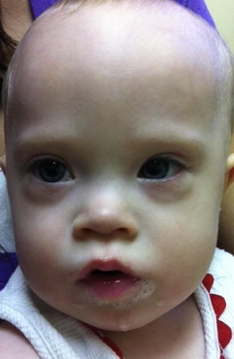
A syndrome is a collection of features that occur together and make up a characteristic clinical entity. In 1886, John Landon Down described the syndrome including microgenia (round face), macroglossia, epicanthal folds, upslanting palpebral fissures, shorter limbs, a single transverse palmar crease, poor muscle tone, mental retardation, and learning disabilities.
The condition was identified as a chromosome 21 trisomy by Jérôme Lejeune in 1959. Today, Down syndrome is the most common congenital chromosome anomaly, occurring in 1 of 700 live births. Recent advances in surgery for the treatment of congenital heart defects have greatly enhanced the survival of children with Down syndrome. Although life expectancy among persons with Down syndrome remains decreased relative to the general population, studies in developed countries document sizable gains in child survival, from 25 years in 1983 to an estimated life expectancy of 50 to 60 years today. As more of these patients are living into adulthood, attention has been focused on health factors that affect the quality of patients’ lives, and affect their ability to reach full potential. A survey of parents attending a Down Syndrome Association conference showed that 50% of children with Down syndrome saw an otolaryngologist regularly. It is likely that the practicing otolaryngologist will encounter many children with Down syndrome, and appropriate treatment can have a significant impact on the quality of life of these patients.
Patients with Down syndrome have several morphologic abnormalities that predispose them to problems with the ear, nose and throat. They have midface hypoplasia with malformation of the eustachian tube, leading to an increased number of ear infections and hearing loss, much of which is preventable with aggressive intervention. They also have shortened palate, relative macroglossia, narrowing of the oropharynx and nasopharynx, and generalized hypotonia, which greatly increases the frequency and severity of obstructive sleep apnea in this population.
Patients with Down syndrome also have alterations in the paranasal sinuses, abnormalities of serum immunoglobulins, and ciliary dyskinesia, which contribute to the high incidence of chronic sinusitis.
In addition, these patients have many comorbidities that must be considered by the surgeon and anesthesiologist:
- •
Congenital heart disease
- •
Pulmonary hypertension
- •
Gastroesophageal reflux disease (GERD)
- •
Subglottic stenosis
- •
Cervical instability.
As care for patients with Down syndrome has been deinstitutionalized in recent decades, and as more of these children are cared for by their parents, integration into schools and social acceptance of these patients has grown. Because of this, more resources have been made available to help these children integrate into society and lead fulfilled, productive, and independent lives. The role of the otolaryngologist in these patients’ lives can be significant. Although most needed in childhood, the otolaryngologist plays a role throughout the life of the patient with Down syndrome ( Fig. 1 ).
Prenatal findings in Down syndrome
On rare occasion, the otolaryngologist may be consulted because of a lack of nasal bones as seen on prenatal ultrasound. A total of approximately 49,000 fetuses from several studies yields a prevalence of nasal bone absence of 65% in trisomy 21 and 1% to 3% in euploid fetuses. If nasal bone evaluation is to be used in screening for Down syndrome, it is important to have sonographers who are trained to perform such evaluations. Differences in studies have ranged from a reported rate of 16.7% for absent nasal bones in fetuses with trisomy 21 evaluated by sonographers without training or quality assurance, to 70% prevalence in studies in which sonographers were appropriately trained.
In the first trimester, the purpose of an ultrasound evaluation of the nasal bones is to recognize whether the nasal bones are present or absent. Ultrasound assessment of nasal bones, in addition to several other ultrasound markers, can increase the performance of first-trimester screening tests to greater than 90% for a fixed false-positive rate of 5%.
In the second trimester, the screening focuses on nasal bone length, with hypoplastic nasal bones being the marker for Down syndrome. Hypoplastic nasal bones are defined as absent or shorter than 2.5 mm. It has been shown that using sonographic markers of prenasal thickness, nasal bone length, and nuchal skin fold increased the detection of Down syndrome in the second trimester by 19% to 23% compared with serum markers alone. This rate of 93% detection for a fixed false-positive rate of 5% is comparable with first-trimester screening protocols. Although absent nasal bones are an important additional finding in prenatal screening, the significance of an isolated finding of absent nasal bones is less clear. One study showed that, in 14 patients with absent nasal bones, only 6 had Down syndrome and 8 had a normal karyotype. In this study, 6 of 6, 100% of fetuses with the isolated finding of absent nasal bones had normal karyotypes; however, 6 of 8 (75%) of the patients who had absent nasal bones in addition to other abnormal ultrasound findings did have Down syndrome. Although there is no role for otolaryngologic intervention at this time, it is helpful for otolaryngologists to be familiar with this screening tool, because they may be consulted for an opinion or assessment.
The ear in Down syndrome
The ears of the patient with Down syndrome are likely the reason for the first encounter with the otolaryngologist. Patients with Down syndrome have a range of otologic problems including stenotic ear canals, increased incidence of secretory otitis media, chronic ear disease, and secondary hearing loss, as well as hearing loss caused by ossicular abnormalities and inner ear dysplasia. In early life, it has been estimated that 40% to 50% of newborns with Down syndrome have stenotic ear canals. These factors may make it difficult for the pediatrician to adequately examine the ear by otoscopic examination.
P earls and P itfalls : These narrowed canals predispose the patient with Down syndrome to cerumen impaction, and the cerumen combined with the stenotic canal makes it difficult to adequately examine the ear. Further, it has been observed that patients with stenotic ear canals had a markedly increased incidence of associated middle ear effusions. It is recommended that children with Down syndrome establish care with an otolaryngologist early in life, because the patient will frequently require microscopic examinations and cerumen disimpaction of the canals under microscopy. It is also recommended that those with canal stenosis continue to follow up every 3 months with the otolaryngologist for evaluation of the middle ear space, to ensure that there is no cerumen impaction, and to monitor for middle ear fluid and infection ( Fig. 2 ).
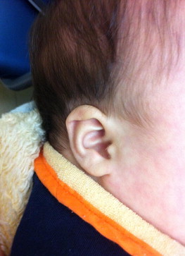
Further research is needed on the natural progression of canal stenosis; however, the experience reported by Cincinnati Children’s Hospital is that most children with stenotic canals grow with age, and by years 2 or 3 this canal is no longer an obstacle to accurate examination. Each patient should be followed regularly by an otolaryngologist until it is clear that the patients are of appropriate age and size so that they are at low risk of serous otitis media and can be easily examined.
Cause of Ear Problems in Children with Down Syndrome
The high prevalence of serous otitis media in children with Down syndrome has been well documented in the academic literature. Several causal factors explain this increased incidence of ear disease:
- •
Children with Down syndrome have an increased number of upper respiratory infections, possibly caused by the reduction of both T and B lymphocyte function, impaired body response to specific pathogens, and defective neutrophil chemotaxis.
- •
Patients with Down syndrome also have midface anatomy that predisposes them to chronic ear disease. The midface hypoplasia seen in many patients with Down syndrome involves the nasopharynx and the eustachian tube openings.
- •
A study on the radiographic features of patients with Down syndrome showed that the bony confines of the nasopharynx were smaller in children with Down syndrome, and therefore the normal-sized soft tissue of the nasopharynx can only occupy this space at the expense of the airway. This decrease in postnasal space may cause even small to medium-sized adenoids to give rise to eustachian tube dysfunction.
- •
It has also been shown that the eustachian tubes in these patients are extremely small, and collapsed in several portions. A histopathologic study of eustachian tubes in patients with Down syndrome compared with controls showed that the cartilage cell density in individuals with Down syndrome was decreased at all ages, predisposing the canal to collapse. Further, it has been hypothesized that the generalized hypotonia of these patients can lead to decreased function of the tensor veli palatini muscle of the palate, which is responsible for opening the eustachian tube. These factors combine to cause collapse of the eustachian tube. This collapse can cause negative pressure in the middle ear space and result in chronic otitis media and fluid accumulation.
- •
In addition to the eustachian tube dysfunction, there may be factors within the middle ear that contribute to disease. In a study of otitis media, Ts65Dn mice, which share many phenotypic characteristics of human Down syndrome, were used as a model for human Down syndrome. When examined, 11 of 15 of the Ts65Dn mice had middle ear effusions, compared with only 2 of 11 of the wild-type mice. On histopathologic examination, the Ts65Dn mice had thickened mucosa and goblet cells were distributed with higher density in the epithelium of the middle ear cavity. Also, bacteria of pathogenic importance to humans were identified in these mice. If these characteristics are also present in the child with Down syndrome, these children are likely to have recurring ear infections.
The causal conditions of secretory otitis media are many, and the impact on the hearing of a child with Down syndrome can be profound. In one study, 90% of children had at least a mild-moderate hearing loss. Despite the high point prevalence of hearing loss in this sample group, only a small percentage of parents (15.2%) reported a positive history of hearing loss.
Hearing Loss in Down Syndrome
It has been documented that hearing loss in children with Down syndrome is more frequent than in healthy children. In studies that conducted audio screening on randomly selected children with Down syndrome, 50% to 90% of children with Down syndrome had a hearing impairment. Hearing impairment may be masked in patients with intellectual impairment, because speech delays, and lack of response to verbal cues, may be attributed to mental retardation.
P earls and P itfalls : It is particularly important for the otolaryngologist to monitor these patients because they are especially susceptible to otitis media and its resulting conductive hearing loss.
Detection of this loss is critical, because the detrimental effects of hearing loss on language development are thought to be greater for children with learning disabilities compared with children without mental retardation. The American Academy of Pediatrics Committee on Genetic and the Down Syndrome Interest Group Guideline recommends audiologic testing at birth, then every 6 months up to age 3 years, with annual testing after 3 years of age, or when ear-specific pure tone audiometry may be obtained ( Fig. 3 ).
P earls and P itfalls : It is recommended that all children with Down syndrome have routine audiologic screening.
Audiologic Evaluation for Children with Down Syndrome
The initial audiologic evaluation is done by auditory brainstem response (ABR) or otoacoustic emissions (OAE). As the patient grows older, the preferred method of audiologic testing is debated.
The evaluation of hearing by behavioral audiometry in young patients is frequently challenging, and is made more difficult in patients who are intellectually challenged. The patient with Down syndrome may have a limited voluntary response during testing, because of a lack of attention and poor psychomotor skills. Further, this type of sound-field testing does not differentiate between ears, because it evaluates both ears together, so a unilateral hearing loss can be missed.
It was shown in a Chinese study that combined objective testing using tympanometry and transitory evoked otoacoustic emission (TEOAE) is a feasible protocol for screening school-aged children with Down syndrome. The TEOAE is able to detect mild hearing loss, but may be inaccurate in the presence of middle ear fluid. The tympanometry and pneumatic otoscopy by the otolaryngologist compliments the examination to screen for hearing loss. However, the TEOAE is costly, limiting accessibility to some patients. Because of cost restrictions, most patients with Down syndrome go for biannual testing using behavioral audiometry, which should be performed by an audiologist who is experienced in behavioral testing and familiar with patients with Down syndrome.
Conductive loss caused by effusion or otitis media
Tympanostomy in Patients with Down Syndrome
The benefits of PE tubes in patients with Down syndrome has been debated during the past decade. The clinical efficacy of tympanostomy tubes has been well established for OME in the general population, with reports that tympanostomy tubes decrease the duration of middle ear effusion compared with no surgical intervention. However, in the patient with Down syndrome, results have been varied.
One study examined the short-term effects of PE tubes, measured by pure tone audiometry, 6 to 9 weeks after tube placement. This study found improved hearing in only 60% of patients with Down syndrome, compared with 91% improvement in the control group. In this study, all patients were older than 6 years (mean age 8.1 years), and none had had PE tubes placed in the past. In the Down syndrome population, which is particularly vulnerable to early onset and prolonged duration of secretary otitis media, this delay of treatment could be the cause of failure, rather than the treatment itself.
A study in Japan also showed poor outcomes in children with Down syndrome, and a high complication rate. This study found that few (21.4%) children were cured of OME by age 7 years, after the initial set of PE tubes was placed, whereas nearly 88% of controls were cured in this same time frame. Reviewing this study revealed faults that do not necessarily indicate treatment failure:
- •
The mean age of these children was 5.4 years, which may be past the critical point in intervening in the Down syndrome population.
- •
The important finding in this study was that most children with Down syndrome had recurrent OME after extrusion of tubes. In normal controls, the incidence of OME is reported to peak in infancy and decline rapidly after the age of 6 years, when the immune system and eustachian tube have reached maturity. However, it has been shown that OME persists for a longer period in children with Down syndrome, and that the canal stenosis, and the highly viscous nature of the mucous expressed, often require further insertion of tympanostomy tubes. The population studied by Iino and colleagues may have had greater success had tubes been reinserted in all patients.
- •
There was a high rate of complications in this study in the patients with Down syndrome compared with controls. Sequelae of OME were found in 15 of 50 of the ears in the Down syndrome group including 4 cholesteatomas, 9 permanent perforations, and 2 atelactatic tympanic membranes. This complication rate has not been reported in other studies; however, it is important to consider when deciding between conservative management and surgical intervention.
Special Considerations in Surgical Management of the Child with Down Syndrome
It is clear from the aforementioned literature that the patient with Down syndrome cannot be treated with the same timeline and same algorithms that are used for the nonsyndromic child. If surgical management is the chosen path, results must be closely monitored, and the surgeon must be aggressive with reintervention. This has been shown by a study at the Cincinnati Children’s Hospital :
- •
Children with Down syndrome were enrolled before the age of 2 years
- •
The children were followed by an otolaryngologist every 3 to 6 months, depending on the degree of canal stenosis present
- •
All children were treated for chronic ear infections and middle ear effusions by placement of PE tubes, and replacement tubes as needed
- •
At the end of the study 2 years later, 93% of the patients had normal hearing
- •
The children with Down syndrome with the PE tubes in place had a 3.6 times higher chance of having normal hearing compared with the audiograms of children with Down syndrome who presented at a similar age as the children finishing the study, but had not had aggressive management of OME and did not have PE tubes in place.
Also reinforcing this concept is a study on receptive and expressive language in adolescents with Down syndrome. In this study:
- •
Adolescents who had tympanostomy tubes placed when they were children showed significantly higher language scores than did the group of patients who had more than 3 known infections as a child, but never had tubes placed
- •
Effects of temporary hearing loss associated with otitis media may play an important role in the language deficits so commonly seen in individuals with Down syndrome
- •
Effects of the hearing loss extend far beyond the time course of the disease itself.
Surgical intervention by placement of PE tubes can be an effective strategy in the management of patients with Down syndrome with OME refractory to medical therapy. However, the otolaryngologist must counsel the parents of the patient about the possible increased risks in these patients, including cholesteatoma, persistent perforation, and atelectatic tympanic membranes.
The study by Iino and colleagues , had a nearly 30% complication rate, which they attribute in part to the eardrums of children with Down syndrome being thin, which can be seen while applying force to the drum with the pneumatic otoscope or while performing myringotomy. They state that a thin eardrum lacks a lamina propria, which lack blood vessels and collagen fibers and are susceptible to permanent perforation. Parents must be prepared for the possibility that the patient may have persistent otorrhea after tube placement. Also, parents must understand that the treatment of OME in the child with Down syndrome may differ from the treatment they may be familiar with or have experienced through other children. They should know that the PE tubes may be placed earlier in the child’s life, and should expect that the child may need multiple set of tubes throughout childhood, even into adulthood. They should be counseled that reinsertion of tubes is a continuation of treatment, rather than failure of the original attempt. Parents should be counseled on the importance of follow-up with the audiologist and the otolaryngologist, and the need for aggressive intervention and reintervention in order for the procedure to be successful at preventing hearing loss.
Conductive loss caused by mastoid or ossicular chain abnormalities
In addition to middle ear effusions, a component of conductive loss may be caused by abnormalities of the mastoid, or abnormality of the ossicular chain. A study reviewed neuroimaging of 59 patients with Down syndrome and found nonaeration or underaeration of mastoids in most (74%) cases. This finding agrees with a previous study, showing 63% of the mastoids examined on lateral cervical spine films with sclerosis and poor aeration. Whether this increase in density is caused by a mastoid infection that occurred during maximum growth years or a congenital component is yet to be determined. A study examining 107 patients with Down syndrome found that only 60% of the conductive hearing loss could be explained by middle ear effusions or tympanic membrane perforations. This finding prompted the investigators to examine the temporal bones of 5 Down syndrome cadavers, as well as document operating room findings during middle ear surgery in these patients. Ossicular abnormalities were attributed to chronic disease, including erosion of the long process of the incus, of the manubrium of the malleus, and of the superstructure of the stapes. Some findings were also attributed to congenital deformities, including malformation of the stapes and dehiscence of the facial nerve. These findings should be considered in children who have a persistent conductive hearing loss despite maximal management of middle ear effusion.
The increased incidence of chronic ear disease in patients with Down syndrome predisposes them to cholesteatoma and erosion of the ossicular chain. Although the 1979 study by Balkany and colleagues found little improvement in conductive hearing loss in patients with Down syndrome who underwent reconstructive surgery for ossicular chain abnormalities, a more recent study by O’Malley and colleagues in 2007 gives more promising results. This study is the largest to date on ear surgery for chronic conditions, including 21 patients with Down syndrome, among 22 other patients with congenital syndromes. This study showed that such patients, including those with Down syndrome, can successfully eradicate disease with creation of a safe, dry ear, and that ossicular chain reconstruction techniques significantly improved hearing in this population. Parents should be counseled that resolution of disease may require several operations. In the this study, 64% of ears were managed with a single surgery, and 89% of ears were controlled with 2 surgeries or fewer. Similarly, parents should also be counseled that canal wall preservation techniques may not be appropriate for this population. In patients with Down syndrome with cholesteatoma in this study, 70% of patients required a canal wall down study, and 89% of patients required a canal wall down procedure in a study of 9 patients with Down syndrome. These procedures have been successful in eradication of disease in this population, but parents should be counseled appropriately about the likelihood of reoperation and the difficulty of using canal wall sparing procedures.
Mixed and sensorineural hearing loss in children with Down syndrome
In addition to the conductive hearing loss caused by otitis media and middle ear effusion, children with Down syndrome also have higher rates of mixed hearing loss and sensorineural hearing loss compared with other children. It is difficult to determine what percentage of this is caused by chronic middle ear disease and osteoid deposition in the fundus of the internal auditory canal and the region of the spiral tract, through which nerve bundles cross from the inner ear to the internal auditory canal. The true incidence of sensorineural hearing loss in children with Down syndrome will be determined as future studies evaluate hearing in children who have been aided by early and aggressive care of their middle ear disease. Although the exact figure of sensorineural hearing loss is difficult to determine, studies evaluating hearing in patients with Down syndrome have found it to be 4% to 9%. Several studies have examined inner ear anomalies that may contribute to hearing loss. One study found them to have uniformly small inner ear structures compared with controls, including hypoplastic cochlea, critically smaller cochlear nerve canal, narrowed internal auditory canal, hypoplastic lateral semicircular canal with a small bony island, and hypoplastic vestibules.
Options to Enhance Hearing of Patients with Down Syndrome
Bone-anchored hearing aid
Early reports on other options to enhance the hearing of patients with Down syndrome are promising. The bone-anchored hearing aid (BAHA) has been successfully used in patients with Down syndrome who failed conventional hearing aids and ventilation tubes. A review of BAHA centers in Ireland and the United Kingdom showed that 18 of 81 BAHA centers are performing surgery on patients with Down syndrome. Of the 43 patients with Down syndrome who were implanted, all but 1 wore the BAHA on a daily basis, which indicates a high level of patient satisfaction.
A survey on perceived patient and parent/caregiver satisfaction was completed by the centers, and showed that 27 of 28 were very pleased or pleased with the results. Similarly, a study surveying 15 patients with BAHA found all 15 using the BAHA regularly, with audiologic benefit.
To evaluate the overall benefit of the BAHA, the Glasgow Children’s Benefit Inventory was used, which evaluates emotion, physical health, learning, and vitality. The results of this study showed a significant benefit in all categories in children with Down syndrome.
Complications in the first study were 50%, which is significantly higher than the previously reported 9% to 16% in nonsyndromic children and 32% in adults. The complication rate in the second study was 20%. In both, the most common complications were soft tissue problems, including excessive healing of the graft site with hypertrophy of soft tissues, graft infection, and skin reaction. All were resolved within a short time, usually within 2 months. The increase in soft tissue complications in patients with Down syndrome may be attributed to patients with learning difficulties having a tendency to interfere with the area, leading to disturbances of the dressing, sutures, and possible graft failure. A solution to this was proposed, in which, following BAHA abutment, a perforated thermoplastic cage is formed over the surgical site and sutured into place. The empirical evidence within this practice has shown this to be effective.
Cochlear implants
Cochlear implants were originally not recommended for patients with additional disabilities beyond hearing loss. However, with a growing body of knowledge and good results, inclusion criteria are expanding and there are now increasing numbers of such candidates, including patients with Down syndrome.
A study published in 2010 reported that at least 4 patients with Down syndrome had received cochlear implants in the United Kingdom and Ireland. In all cases, the deafness was congenital, and all 4 of the patients had middle ear disease before surgery, with 2 patients requiring PE tubes. However, all of the patients were treated before surgery and none of the cases had any complications associated with otitis media. Despite the previous discussion of mastoid underaeration and opacification, inner ear dysplasia, shortened cochlear lengths, and dehiscent facial nerve, none of the patients implanted had any of these findings and none had intraoperative difficulties.
The outcomes for these 4 implanted patients have been modest gains in auditory performance, with the eldest child, who has had the implant the longest, showing the most improvement. As more patients with Down syndrome become candidates for cochlear implants, patients and families must be counseled about expectations. There are abnormalities in the temporal bone of a child with Down syndrome that may increase the risk of complications. Even in technically successful implantation, the outcomes may not be as good as in children without additional disabilities, because learning and communication difficulties may prolong the rehabilitation. However, these patients do show improvement, and future patients with Down syndrome with profound sensorineural hearing loss may be referred for assessment at cochlear implant programs.
Sound-field amplification and speech/language intervention
In addition to surgical options, initial studies using sound-field amplification and speech/language intervention have shown excellent results.
A pilot study tested an aggressive multidisciplinary model consisting of amplification technology and speech/language intervention that emphasizes auditory-verbal therapy, as well as aggressive medical and surgical management of ear disorders. This program was initiated in children with Down syndrome less than 1 year old. The findings in these 6 children enrolled in the program were that the children in the intervention group had developed age-appropriate early language skills, with no apparent gap between their receptive and oral expressive language abilities. A group of children with Down syndrome of the same age who did not have the intervention were used as comparison, and the no-intervention group had generalized language delays with a noticeable gap between receptive understanding and oral expressive language.
Another study examined the benefits of sound-field amplification in 4 children with Down syndrome in the classroom setting. The study found that that participant’s speech perception significantly improved when the FM sound-field amplification was being used. The sound-field amplifier is recommended rather than a traditional hearing aid in this population, because the sound-field amplifier selectively amplifies the teacher’s voice, which improves the signal/noise ratio, whereas the hearing aid increases all sounds equally, including background noise that can be distracting. In the patients with Down syndrome, who are prone to fluctuating conductive hearing loss, the effects of poor classroom acoustics are significant.
As more of these children with Down syndrome are mainstreamed and placed in public schools, additional support is needed to achieve full potential. More outcomes research is needed, but sound-field amplifiers have the potential to improve classroom performance.
Obstructive sleep apnea and sleep disordered breathing
Although OSAS is seen in only 0.7% to 2.0% of the general pediatric population, the prevalence in pediatric patients with Down syndrome has been estimated at 77% to 80%. Children with Down syndrome have many predisposing factors of OSAS:
- •
Midfacial and mandibular hypoplasia glossoptosis
- •
An abnormally small upper airway with superficially positioned tonsils and relative tonsillar and adenoidal encroachment
- •
Increased secretions
- •
Increased incidence of lower respiratory tract anomalies
- •
Obesity and generalized hypotonia with resultant collapse of the airway during inspiration.
Children with OSAS have a worsened trend in word reading speed, visual attention, and verbal fluency. OSAS has been shown to result in neurodevelopmental problems such as daytime somnolence, behavioral disturbances, school failure, and developmental delay. Obstructive sleep apnea can cause pulmonary hypertension resulting in cor pulmonale and congestive heart failure secondary to the chronic, intermittent hypoxemia and respiratory acidosis during sleep. Although we know of no published studies specifically examining these effects of neurodevelopment and learning on children with Down syndrome, it is logical to expect that this population, already predisposed to learning delay and difficulty in school, would be significantly impaired by the effects of sleep apnea. Similarly, children with Down syndrome are predisposed to congenital cardiac anomalies and are more likely to have pulmonary hypertension than are nonsyndromic children with the same cardiac anomalies. Again, this may be exacerbated or worsened by OSAS ( Fig. 4 ).
P earls and P itfalls : Children with Down syndrome have many predisposing factors of OSAS.
Diagnosing Obstructive Sleep Apnea in Children with Down Syndrome
The diagnosis of OSAS should come from an overnight polysomnography whenever possible. A study examined 53 patients with Down syndrome for OSAS by nap study and, of those, 16 patients had both a nap polysomnography and an overnight polysomnography. All 16 (100%) had abnormal overnight polysomnograms, but the nap study was less sensitive in detecting OSAS, with only 12 (75%) of these patients having abnormal nap studies. The degrees of hypoventilation and desaturations were significantly higher in the overnight studies, and thus the nap studies, under estimated abnormalities.
Snoring in sleep apnea diagnosis
The diagnosis of sleep apnea is not limited to the children with Down syndrome who snore, although those who do snore have a high likelihood of having a positive sleep study. In a retrospective review of patients with Down syndrome who were referred for polysomnography because of snoring, 97% of these snoring patients had a positive sleep study. The child with Down syndrome who is not reported to snore is still at significant risk for sleep apnea and should be evaluated.
Parental reporting of sleep apnea
It may be difficult for a parent to tell whether a child is suffering from sleep apnea, because the most severe apneic events often happen during rapid eye movement (REM) sleep, late at night, when the parents are also asleep. In the child with Down syndrome, the parents may assume that their child’s irregular breathing at night is normal for a child with Down syndrome, which is a frequently expressed comment. Parental reports are not reliable in ruling out sleep apnea.
A study showed that 11 of 35 (31%) of parents of children with Down syndrome reported that their children had sleep problems, but these parents were correct about a sleep abnormality in only about 4 of 11 (36%) cases. The other 7 of 11 (64%) had normal polysomnograms. Of the 24 of 35 (69%) parents who reported no sleep problems, 13 of 24 (54%) of the children had abnormal polysomnograms, and did have obstructive sleep apnea.
In another study, 19 of 49 children (39%) had histories that suggested OSAS. Polysomnograms were abnormal in all 19 (100%) of the patients with a positive history. However, 18 of 30 (60%) of the children with negative histories also had abnormal polysomnograms.
Because of the unreliability of parental reporting, the high prevalence of OSAS in this population, and the negative effects of sleep apnea, it is recommended that all children with Down syndrome between the ages of 3 and 4 years go for objective testing using full overnight polysomnography for a baseline study.
Risk Factors for OSAS
Because of the possibility of health problems associated with OSAS in a population already at higher health risk, learning about the cause of the disease is important.
One established risk factor for obstructive sleep apnea (OSA) in adults and children in the general population is high body mass index (BMI), and weight reduction is often effective at decreasing the effects of sleep apnea. However, the correlation between BMI and OSAS in the patient with Down syndrome is not so clear.
- •
A study surveyed consecutively encountered, nonselected patients with Down syndrome. Seventy-nine percent of them had OSAS, and higher BMI was significantly associated with a higher apnea index and lower arterial oxygen saturation (Sa o 2 ) level.
- •
A study in 2010 compared age-matched children with Down syndrome with OSA and without OSA based on polysomnogram results. The mean BMI z-score was statistically significantly different between OSA and non-OSA groups, with the OSAS group having a BMI z-score of 2.09 and the non–sleep apnea group a BMI z-score of 1.4. The BMI z-score, also called BMI standard deviation score, are measures of relative weight adjusted for the child’s age and sex. However, there were some patients in this review who had an extremely high BMI who had a normal sleep study, and several patients with a low BMI did have OSAS.
- •
Similarly, in the study by Fitzgerald and colleagues in 2007, 91% of the study subjects were not obese, but 97% had OSA and 50% of those had severe OSA.
Based on these results, OSAS is likely a multifactorial disease with several contributing factors in these patients. However, BMI is a modifiable risk factor, and the results of the studies discussed earlier suggest that weight reduction may show some benefit in the management of OSA in children with Down syndrome ( Fig. 5 ).
P earls and P itfalls : A preoperative polysomnogram is now recommended in patients with Down syndrome before tonsillectomy and/or adenoidecomty.