, Maneli Mozaffarieh1 and Hans Bebie2
(1)
Department of Ophthalmology, University of Basel, Basel, Switzerland
(2)
Institute for Theoretical Physics, University of Bern, Bern, Switzerland
Abstract
What happens when light meets matter? There is always an interaction: light is scattered at a wall’s surface, reflected off a surface of water, partially absorbed and partially reflected by a green leaf, refracted when it enters glass, and excites chemical processes in retinal rods and cones, even at very low intensities. The details depend on the structure of the matter and on the wavelength of the light. Additional phenomena are refraction, diffraction, and fluorescence – even the miracle of transparency is fascinating. How is it possible that light passes almost completely unimpeded through a structure like the cornea or through water molecules? In this chapter, we discuss how light is affected by matter. In Chap. 7, we will discuss the special action of light on tissues.
What happens when light meets matter? There is always an interaction: light is scattered at a wall’s surface, reflected off a surface of water, partially absorbed and partially reflected by a green leaf, refracted when it enters glass, and excites chemical processes in retinal rods and cones, even at very low intensities. The details depend on the structure of the matter and on the wavelength of the light. Additional phenomena are refraction, diffraction, and fluorescence – even the miracle of transparency is fascinating. How is it possible that light passes almost completely unimpeded through a structure like the cornea or through water molecules? In this chapter, we discuss how light is affected by matter. In Chap. 7, we will discuss the special action of light on tissues.
2.1 Phenomenology
Almost all of the processes mentioned above can be illustrated using the eye as an example. Thanks to the refraction of light at the air–corneal interface and at the aqueous humor–lens interfaces, a sharp image is engendered on the retina. The cornea reflects a crossbar or a Placido disk. The aged lens scatters light and reduces the image contrast at the level of the retina. Blood mainly absorbs blue and green light and converts the energy into heat so that red is the dominant color in the light that is scattered back.
The blue iris owes its color to the same process that produces a blue sky: i.e., light scattered by particles that are smaller than the light wavelength. Shorter wavelengths are scattered much more than the longer ones (Rayleigh scattering). The color of a brown iris arises from absorption by a pigment. The white color of the sclera is explained by the almost total scattering of all colors in every direction. In fluorescence angiography, the conversion of light to longer wavelengths is applied. Due to its wave properties, even the diffraction of light is manifest within the eye: the smaller the pupil is, the larger the smallest image of a point source of light at the retina will be. A few of the more important processes are depicted in Figs. 2.1 and 2.2. In the following chapters, we discuss in detail some of these processes and their ocular manifestations.
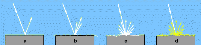

Fig. 2.1
Some of the interactions of white light with surfaces. (a) Specular reflection at a smooth surface. (b) Lustrous reflection from paper with a slightly rough surface. (c) Diffuse reflection from a whitewashed wall; no absorption. (d) Diffuse reflection with absorption of the shorter wavelengths at a painted yellow wall
Fig. 2.2
Some interactions of white light with media. Refraction takes place when a ray of light penetrates from above into the medium below (as seen in these diagrams). The media are, for example, gases, fluids, or tissues. (a) Refraction. In the denser (lower) medium, light travels more slowly and in a changed direction. (b) Scattering, not color-selective (strongly diluted milk). (c) Absorption without scattering (clear medium). After blue has been absorbed, the remaining ray of light is yellow. (d) Absorption of blue and green, additional scattering of the light (cloudy medium such as blood)
2.2 Fundamental Physical Processes
We shall occupy ourselves only briefly with the atomic bases of the mentioned processes. The basic principle is always the same with visible, ultraviolet, or infrared light. When light encounters a surface or passes through a medium, inevitable interaction occurs between the light and the electrons of the atoms and molecules of the material. A simplified picture of classical electrodynamics involves the interaction of two fundamental processes: first, the light exerts a force on the electron1 and, second, as a charged particle, the accelerated electron radiates electromagnetic waves (light).
Scattering of light by a free electron provides an example. When light meets an electron, it is “shaken” at the frequency of the light. As a result, the electron sends out light with the same frequency in any direction. Thus, light scattering takes place. This process represents one of the impediments that solar photons surmount when they must fight their way from where they are produced in the interior of the sun to its outer surface. A second example is that light penetrating through a metallic surface causes the cloud of negative charge – consisting of weakly bound electrons of the metal atoms – to vibrate in phase with the light frequency. This vibrating and charged cloud then produces light of the same frequency, specifically reflected light. A third example is that, inside glass, electrons are also stimulated to vibrate. Instead of reflection, the only consequence in glass is that the light is slowed down somewhat without being absorbed. This slowing down of the light is the basis for refraction (Sect. 2.4).
The electric field of light exerts forces of the same strength on the protons of the atomic nuclei as it does on the electrons. However, due to the much larger mass of the protons and their strong binding within the atom’s nucleus, the interaction of visible light with the nucleus is far weaker and is practically negligible in the visible range.
The basic process of the interaction of light with matter can be described more precisely by means of quantum theory: the electron of an atom, a molecule, or an atomic lattice can absorb a photon and use its energy to jump into an energetically higher state (Fig. 2.3). Conversely, an electron can fall into a state of lower energy, with the energy difference being sent out as a photon (Fig. 2.4). Actually, it is usually not just a single electron but, rather, the whole shell of an atom or molecule that experiences a change of state in these processes. Besides these two basic processes (absorption and emission), there is a third one: stimulated emission. This will be treated in Sect.3.4.
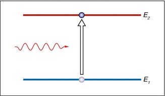
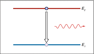
Fig. 2.3
Absorption of a photon. Its energy is transferred to the atom and raises its electron shell onto a higher energetic state. This process is only possible when the photon’s energy “fits” a gap in the atom’s energy spectrum
Fig. 2.4
Spontaneous emission of a photon by an excited atom or molecule. Typically, this process occurs spontaneously, often only a few nanoseconds after the absorption of energy. The energy difference between the two atomic states determines the frequency (and, thus, the wavelength) of the departing photon. The direction of flight of the emitted photon is random
Scattering and absorption fit quite simply into this picture of elementary processes. Scattering means that a photon is absorbed and immediately emitted again. The absorbed energy equals the emitted energy and, as a result, it does not change the wavelength of the light. The absorption of light by a black piece of paper or by the pigments of a brown iris follows another scheme: first, the absorption of a photon results in the transition of an atom or molecule to a state of higher energy. This energy is now converted in small portions into vibrations of the material. Heat is generated from the photon’s energy (Fig. 2.5). Which one of the aforementioned processes takes place depends on the material, more precisely on its structure and molecular composition.
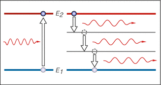
Fig. 2.5
Absorption of a photon and dispersion of the energy into lattice vibrations. The absorbed light energy warms the absorber
2.3 Transparency
Keeping in mind that light is scattered when it encounters an obstacle, the existence of transparent media such as glass, water, corneas, crystalline lenses, and air seems quite miraculous. Inside these media, interactions between the light and the materials still occur, but it only leads to the light’s traveling more slowly than it would in a vacuum.2 This slowing down is quantified as the refractive index n: the velocity of light in the medium amounts to c′ = c/n, where c is the velocity of light in vacuum (c ≈ 300,000 km/s). For example, in water, light travels with a velocity of c′ ≈ 225,000 km/s (n = 1.33).
Pure water is a classic example of an almost completely transparent medium for visible light. An eye exhibits several portions of tissue that are more or less transparent, such as the cornea, crystalline lens, aqueous humor, and the vitreous body, as well as the inner layers of the retina. A medium is always transparent only to a certain part of the electromagnetic spectrum. For example, water is opaque to radiation in the infrared range (see Sect. 2.8), while the cornea blocks radiation in the ultraviolet range.
It is impressive how nature has been able to construct transparent tissues. The cornea is made up of multiple layers (Figs. 2.6 and 2.7). The largest portion consists of the so-called stroma, which contains relatively few cells but many collagen fibers. For the stroma to be transparent and remain so, a very special arrangement of these collagen fibers must be maintained. The fibers are packed tightly and run from limbus to limbus. The cornea is transparent only as long as the separation between the collagen fibers is less than half a wavelength of the light that passes through. If too much water is present in the stroma (for example, when the pump function of the endothelium fails), the collagen fiber separation increases and the cornea loses its transparency (Fig. 2.8). This can occur, e.g., in cases of corneal decompensation. Here, we have a situation where the incorporation of clear, transparent water leads to clouding of the corneal medium.
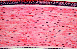
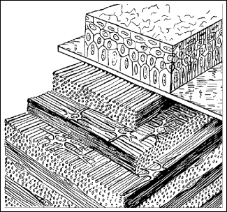
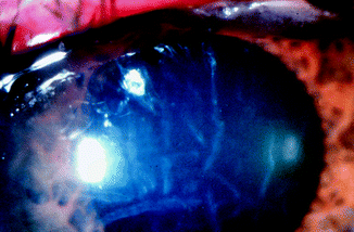
Fig. 2.6
Multi-layer construction of the cornea (Courtesy of E. van der Zypen, University of Bern)
Fig. 2.7
Multi-layer construction of the cornea (Courtesy of H. Elias and J. E. Pauly (1966) Human Microanatomy. F.A. Davis Comp., Philadelphia. With permission)
Fig. 2.8
Reduced corneal transparency due to swelling of the stroma
The crystalline lens of a healthy person is also transparent. It consists of the capsule, the epithelium, and the lens fibers. The lens fibers run in a meridional fashion from the posterior to the anterior poles (Fig. 2.9). Again, the regular arrangement of these fibers is a prerequisite for the transparency of the lens.
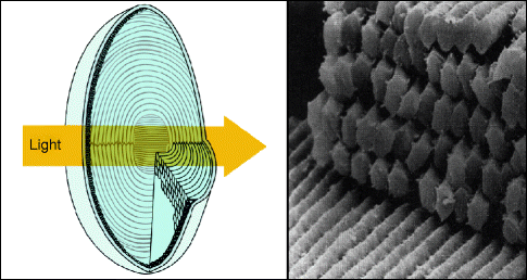
Fig. 2.9
Regular ordering of the lens fibers. Right: Scanning electron micrograph showing the orderly arrangement of hexagonal lens fibers. (Right figure from Adler’s Physiology of the Eye (2003) Mosby. With permission. Courtesy of J. Kuszak)
The retina is also transparent, so light can reach the cones and rods unimpeded (Fig. 2.10). However, it can also lose its transparency through water retention (retinal edema). A similar phenomenon can occur at the optic nerve head. The nerve fiber layer continues from the retina into the optic nerve head. The nerve fiber layer is transparent, so, in ophthalmoscopy, the ophthalmologist sees through this layer to deeper layers and, thereby, sees the clear, sharp boundaries of the retina, pigment epithelium, and choroid (Figs. 2.11 and 2.12). The optically sharp delimitation of the optic nerve head is, thus, conditioned by deeper layers. If the nerve fiber layer loses its transparency, either partially or totally, the optic nerve head’s boundaries appear blurred. This loss of nerve fiber transparency is encountered, for example, in cases of papilloedema in which the axons swell and thereby lose their transparency (Fig. 2.13).
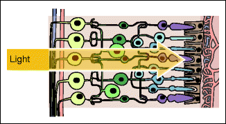
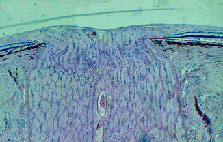
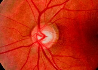
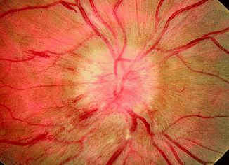
Fig. 2.10
Transparency of the retina
Fig. 2.11
Both the choroid and the pigment epithelium end sharply at the border of the optic nerve head, whereas the superficial nerve fiber layer is continuous (Courtesy of P. Meyer, University of Basel)
Fig. 2.12
Sharply defined retina, pigment epithelium, and choroid
Fig. 2.13
In the case of papilloedema, the nerve fibers lose their transparency. This gives the impression of a blurred-bordered optic nerve head
2.4 Refraction
If a beam of light meets a smooth interface between two transparent media that have different refractive indices, both reflection and refraction occur (Fig. 2.14). The reflection is symmetric with respect to the surface normal,3 and the percentage of the light reflected increases with an increasing angle α. In Sect. 2.5, we will discuss reflection in more detail. The refraction of light is the basis for the optical imaging through the cornea, crystalline lens, eyeglasses, and optical instruments (e.g., magnifying glasses, microscopes, and refractive telescopes).
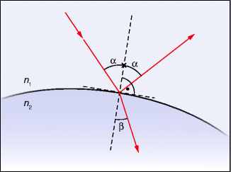
Fig. 2.14
Refraction at the interface of two media. The primary ray is partially reflected and partially refracted. α and β are the angles of the rays with respect to the surface normal. The law of refraction determines the angle β when α and the refractive indices n 1 and n 2 are given: sin α / sin β = n 2 /n 1
2.4.1 The Law of Refraction
The incident ray of light onto a surface, the refracted and reflected rays, and the surface normal all lie in the same plane (Fig. 2.14). The amount of light refracted depends on the ratio of the refractive indices of the two media. The relationship between the two angles α and β is specified by the law of refraction (Fig. 2.14). Depending on which culture one comes from, it is ascribed to either Snell4 or Descartes,5 both of whom rediscovered it at roughly the same time.6
Refraction is a consequence of the differing speeds of light in two media (Fig. 2.15). To understand this, we first note that the frequency of the light vibrations remains the same in both media (the vibrations on both sides of the interface are in step). Therefore, inside the medium with the slower light speed, the wavelength is smaller since the light moves one wavelength further during one period. Figure 2.15 shows that the continuous transition at the interface is possible only with a change in direction.
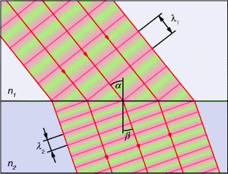
Fig. 2.15
Wave image of refraction. The differing light speeds in the two media give rise to differing wavelengths. The continuous transition of the phases at the interface is possible only with a change in direction. In a medium with an index of refraction n, the wavelength is n times shorter than in a vacuum. λ 1 / λ 2 = n 2 / n 1
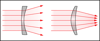
Lenses that are thinner in the center are called diverging lenses (“minus lenses”). Their optical powers are given as negative values. Lenses that are thicker in the center than the periphery are called collecting lenses (“plus lenses”). They bundle parallel light into a focus. The reciprocal value of the focal length (in meters) is called the optical power and is measured in diopters, D (to give an example, a focal length of 0.25 m corresponds to a refractive power of 4 D). Bending a lens inward or outward slightly does not influence its refractive power (Fig. 2.16).
 < div class='tao-gold-member'>


Only gold members can continue reading. Log In or Register to continue
Stay updated, free articles. Join our Telegram channel
Full access? Get Clinical Tree
