Diabetes mellitus (DM) involves a complex sequence of events that often evolve over many years. It is characterized by a broad array of abnormalities and frequently results in serious, long-term vascular and neurologic complications. Pathologic changes may lead to systemic hypertension, renal failure, blindness, autonomic and peripheral neuropathy, amputations of the lower extremities, myocardial infarctions (MIs), peripheral artery disease, and strokes.
The most notable feature of the disease is a disordered metabolism of glucose leading to hyperglycemia. Elevation in blood glucose results from an absolute deficiency of insulin secretion, a reduction in its effectiveness (insulin resistance), or an increased hepatic glucose output. Chronic hyperglycemia is presumed to cause tissue damage by glycation of proteins, by altering cell signaling pathways, and/or excess production of polyol compounds from the metabolism of glucose.
There are two major types of DM: type 1 (formerly juvenile onset, type I or insulin-dependent diabetes mellitus—IDDM) and type 2 (formerly adult onset, type II or noninsulin-dependent diabetes mellitus—NIDDM). Other less common forms of DM exist but are beyond the scope of this section. Each form of DM has a distinct etiology, clinical presentation, and course.
Note: The terms and acronyms IDDM and NIDDM are no longer used since the names are based on pharmacological treatment rather than causative considerations. The current classification system recognizes that DM results from different mechanisms, with the common endpoint being hyperglycemia.
Etiology
Type 1—Type 1 diabetes mellitus is an autoimmune disease in which pathologic, autoreactive T cells attack the insulinsecreting pancreatic islets of Langerhans, thereby leading to lifelong dependence on insulin for affected patients. This disease process often results in the production of autoantibodies, such as anti-islet cell autoantibodies (ICAs), autoantibodies to insulin (IAAs), and glutamic acid decarboxylase (GAD65). There is evidence that a precipitating event, such as a viral illness, may be required to trigger the destructive process. Other studies suggest that toxins in the environment, or diet supplementation with gluten-containing foods before age 3 months, might also initiate damage of genetically vulnerable beta cells. Whatever the etiology, clinical diabetes occurs when roughly 90% of the beta cells have been destroyed; this process can be quite rapid in infants and children and slower in adults. Circulating insulin in type 1 diabetes is virtually absent, thus these patients are dependent on exogenous insulin for survival. Ketosis is common in patients with poor glucose control and ketoacidosis can be life threatening.
Note: The type 1 Diabetes TrialNet studies (http://www.diabetestrialnet.org) are investigating the treatment of patients who have newly diagnosed type 1 diabetes, using immunologic agents (i.e., rituximab, mycophenolate mofetil, and daclizumab) to determine if these drugs can prevent further beta cell destruction. The studies may demonstrate that the incidence of type 1 diabetes can be reduced.
Type 2—The exact etiology of type 2 diabetes mellitus continues to be debated; however, development can often be correlated with obesity, in particular increases in visceral fat. Some patients may initially show normal insulin levels, but the natural history of type 2 diabetes reveals a gradual decrease in the number of insulin-producing beta cells, resulting in a relative insulin deficiency that worsens over time. At the point of diagnosis, a patient may have already lost 50% of their beta cell function, and he or she will typically lose 4% per year thereafter. Rising glucose levels further compromise insulin secretion and insulin action, a phenomenon known as “glucose toxicity.”
Ultimately, patients with type 2 diabetes develop a “dual defect” disease characterized by insulin resistance and a defect in compensatory insulin secretion. These individuals may also exhibit reduced numbers of insulin receptors, increased hepatic glucose production, impaired glycogen synthesis, paradoxical rise of glucagon in response to meals, decreased peripheral glucose uptake, abnormal patterns of insulin secretion, accelerated gastric emptying, or a combination of these conditions. Circulating endogenous insulin is usually sufficient to prevent ketoacidosis but inadequate to prevent hyperglycemia.
Note: It is now believed that about 2 million Americans currently diagnosed with type 2 diabetes may actually have latent autoimmune diabetes in adults (LADA), also referred to as type 1.5 DM. This condition appears to be a hybrid or “stealth” type of diabetes, which progresses slower in its attack and destruction of insulin-producing beta cells than type 1. Patients with LADA cannot normalize their blood glucose with oral antidiabetic agents and thus require exogenous insulin supplementation.
The Patient
Clinical Symptoms
Some cases of DM (especially type 2) are diagnosed at an asymptomatic stage as a result of a screening program, routine blood tests, or an eye examination. Compared with type 1 patients who commonly present with acute symptoms, individuals with type 2 diabetes are much more likely to have no symptoms or fewer symptoms, as the hyperglycemia develops gradually. Patients may report the classic syndrome of polyuria, polydipsia, polyphagia, unexplained weight loss, weakness, and fatigue; however, these symptoms are typically associated with marked hyperglycemia as seen in type 1 patients (Table 16-1).
Clinical Signs
The physical findings will vary depending on the type of DM, the severity of insulin deficiency, and the duration of the disease. Examination may reveal systemic hypertension, neuropathy, periodontal disease, vaginitis, skin abnormalities, arteriopathy, blurred vision, retinopathy (Figs. 16-1 and 16-2), nephropathy, and/or increased susceptibility to infections. Diabetics also have a much higher risk for cardiovascular (CV) disease, abnormalities in lipid metabolism (dyslipidemia), and vascular thrombosis (see Table 16-1).
Ocular manifestations of DM occur frequently and careful examination with appropriate treatment can often reduce the ophthalmic complications (Table 16-2). Clinicians should also be aware that the presence of any diabetic retinopathy (DR) signifies an excess risk of renal failure and of heart failure (HF) and that visual impairment, resulting from DR, predicts a high mortality rate, particularly from CV diseases and stroke.
*Note: While many of these signs and symptoms may occur in both type 1 and type 2 diabetes, those conditions indicated with a dagger
† are most commonly detected in type 1 disease and associated with marked hyperglycemia. Retinopathy may be detected in over 20% of type 2 patients at the time of initial diagnosis and may predate the diagnosis of diabetes by up to 6 years.
Early detection, improved delivery of care, and better self-management are key strategies for preventing diabetes-related complications. (For every 1% decrease in the A1c, there is approximately a 35% reduction in the risk of diabetic microvasculature complications.)
Demographics
The estimated lifetime risk of developing DM for individuals born in 2000 is 32.8% for males and 38.5% for females.
DM is the sixth leading cause of death in the United States and is believed to be underreported both as a condition and as a cause of death. It is estimated that up to 17% of all deaths in the United States are related to diabetes.
The incidence, target population, and genetics differ between type 1 and type 2 (Table 16-3).
It is estimated that over 27 million people in the United States (>200 million worldwide) have DM and that the number of patients with diabetes will double every decade.
About a fourth of U.S. adults, or 57 million people, have prediabetes. These patients have a substantially increased risk of CV disease and death.
Type 1 diabetes affects 1 in 250 persons in the United States. Approximately 10,000 to 15,000 new cases are reported each year, and the disease appears to be on the rise.
Type 1 diabetes is less common among black and Asian populations.
Type 2 diabetes accounts for approximately 90% to 95% of all cases of diabetes among American adults.
Type 2 diabetes has an overall prevalence of approximately 9% in the United States, but it is estimated that by 2010, over 10% of the population will have type 2 diabetes. One and a half million new cases are reported each year; approximately 25% to 30% of patients are unaware of their disorder at the time of diagnosis.
Type 2 diabetes is more common in Native Americans, African Americans, and Hispanic/Latino Americans. This same population has a three- to sixfold higher incidence of diabetes-related end-stage renal disease than seen in whites.
Type 2 diabetes increases with age, lack of physical activity, and obesity. Individuals who have a body mass index (BMI) greater than 30 are at 10 to 20 times greater risk. Approximately 28% of U.S. adults aged 20 years and older are obese.
Type 2 diabetes occurs more frequently in women with prior gestational DM; these women have a 20% to 50% chance of developing diabetes over 5 to 10 years.
DR is the leading cause of new cases of blindness in patients 25 to 74 years of age. In 2007, DR affected more than 5.5 million Americans, and it is predicted that the number of cases will at least triple by 2050.
Each year, 12,000 to 24,000 people lose their sight due to diabetes. African Americans are twice as likely to suffer from diabetes-related blindness.
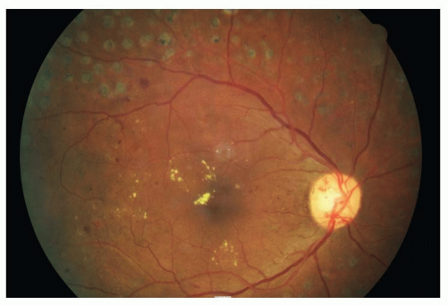
Figure 16-1. Proliferative DR with NVD and CSME. Note prior PRP scars in the periphery.
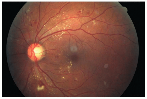
Figure 16-2. Moderate to severe nonproliferative DR with CSME.
TABLE 16-2 Ocular and Periocular Manifestations of DM
Glaucoma
Possibly increased incidence of POAG
Pupils
Light-near dissociation
Refraction
Fluctuations in refractive errors
Cornea
Hypesthesia, corneal abrasions, poor healing
Iris
Neovascularization of the iris or angle. Obtain retinal consult within 2-4 d. PRP may prevent development of neovascular glaucoma
Ectropian uveae
Lens
Premature cataracts
Ciliary body
Premature presbyopia
Extraocular muscles
III, IV, VI nerve palsies (mononeuropathy)
Vitreous
Detachment, hemorrhages
Treat a preretinal and/or vitreous hemorrhage as high-risk proliferative DR. Obtain retinal consult within 24-48 h
Optic nerve
Ischemic optic neuropathy or papillopathy
NVD
Macula
Diabetic maculopathy is the most common cause of visual loss in diabetes and may be exudative, edematous, or ischemic
CSME
Thickening of the retina at or within 500 µm from the center of the macula.
Hard exudates at or within 500 µm from the center of the macula if there is thickening of the adjacent retina.
Zone or zones of retinal thickening, one disc area or larger, any part of which is within one disc diameter of the center of the macula.
CSME may be present at any stage of DR and should be treated with focal or grid photocoagulation. Intravitreal injection of triamcinolone acetonide or anti-VEGF agents show promise for the treatment of diabetic ME that is unresponsive to laser photocoagulation. Obtain retinal consult within a 2-4 wk period
Retina
Mild to moderate nonproliferative retinopathy; no CSME
Follow every 6-12 months depending on extent of lesions
Severe nonproliferative retinopathy (preproliferative); no CSME
Increased intraretinal hemorrhages and exudates, venous beading and loops, IRMA, CWS, large areas of capillary nonperfusion.
Eyes with hemorrhages (>20) in four quadrants, or venous beading in two or more quadrants, or IRMA in one or more quadrant are considered to have severe NPDR. Patients with two or more of these characteristics have very severe NPDR
Obtain retinal consult within 3-4 wk and follow every 2-4 mo
Proliferative retinopathy; no CSME
NVD
Retinal NVE
Preretinal or vitreous hemorrhage
Requires PRP
Obtain retinal consult within 2-4 d if high-risk characteristics are present
Retinal detachments
Tractional
Rhegmatogenous
Obtain retinal consult within 3-5 d
Other vision threatening retinal conditions which are more prevalent in the patient with diabetes include the following:
Branch retinal vein occlusion
Central retinal vein occlusion
Branch retinal artery occlusion
Central retinal artery occlusion
Anterior ischemic optic neuropathy
Diabetic papillopathy
Patients >10 years of age with type 1 diabetes should have an initial dilated and comprehensive eye examination within 3 to 5 years after the onset of diabetes, and at least every year thereafter.
Patients with type 2 diabetes should have an initial dilated and comprehensive eye examination shortly after diagnosis, and at least every year thereafter.
TABLE 16-3 Comparison of the Essential Features of Type 1 and Type 2 DM
Type 1
Type 2
Other names (outdated terms)
IDDM, juvenile onset, growth onset, ketosis-prone, type I
NIDDM, adult onset, maturity onset, ketosis resistant, type II
Age of onset
Usually under 35 but can occur at any age
Usually over 35 but the incidence is rapidly increasing in adolescents
Nutritional status at onset
Usually nonobese
Usually obese
Symptoms
Polyuria, polydipsia, polyphagia
Frequently none
Ketosis
Common, unless diet, insulin, and exercise are coordinated properly
Uncommon except in the presence of infection or stress
Endogenous insulin secretion
Severe deficiency to absent
Moderate deficiency; may be in excess but relatively ineffective due to obesity, that is insulin resistance
Associated lipid abnormalities
Dyslipidemia increases with age
Cholesterol and triglycerides frequently elevated, decreased HDL
Insulin requirements
Always necessary
Needed in 20%-30% of patients
Insulin resistance
Occasional
Almost always
Oral agents
Sometimes efficacious
Frequently efficacious
Diet
Mandatory along with insulin supplementation
Diet alone frequently adequate to control blood glucose
HLA association
DR3, DR4
None
FH of DM in a first degree relative
∼20%
∼70%-80%
Identical twins
∼50% concordant
∼100% concordant
ICAs or insulin autoantibodies (IAAs)
Autoantibodies are detected in 85%-90% of patients
Absent
Association with other autoimmune diseases
Frequent
None
Significant History
Age (type 2 DM is more common in people greater than 45 years of age; however, it is becoming increasingly more common in younger individuals, even adolescents)
Family history (FH) of DM in a first-degree relative; if FH is positive for DM, determine age of onset, whether it was associated with obesity, and form of treatment. FH is much stronger in type 2 (70% to 80%) versus type 1 (20%)
Diabetic patients with family members who have had severe retinopathy are more likely than other diabetics to have more severe retinal disease
Unexplained weight loss (type 1) or visceral obesity and increased BMI (type 2)
Physical inactivity
Polyuria, polydipsia, polyphagia may be reported with marked hyperglycemia
Chronic or recurrent infections
Gestational diabetes, or having delivered a baby weighing greater than 9 lb
Long or highly irregular menstrual cycles
Member of a high-risk ethnic population
Systemic hypertension, smoking, dyslipidemia, use of oral contraceptives, or other factors that increase cardiac risk
Ancillary Tests
The risk of developing type 2 diabetes is increased with plasma glucose (PG) levels well below the current cut-point for defining impaired fasting glucose (IFG), which is 100 to 125 mg/dL. Fasting plasma glucose (FPG) levels, even as low as 90 mg/dL, have been shown to be associated with an increased risk for type 2 diabetes. However, diabetes risk associated with fasting glucose should be viewed in the context of other risk factors such as systemic hypertension, dyslipidemia, and obesity. While the definition or cut-point of DM may change with time, the conventional criteria are as follows:
A diagnosis of DM, in nonpregnant adults, is appropriate in one of the following circumstances. Each must be confirmed on a subsequent day unless unequivocal symptoms of hyperglycemia are present:
Symptoms of diabetes plus casual PG concentration ≥ 200 mg/dL (11.1 mmol/L). Casual is defined as any time of day without regard to time since last meal. The classic symptoms of diabetes include polyuria, polydipsia, and unexplained weight loss.
FPG ≥ 126 mg/dL (7.0 mmol/L). Fasting is defined as no caloric intake for at least 8 hours. If the FPG level is over 126 mg/dL on more than one occasion, further evaluation of the patient with a glucose challenge is unnecessary.
2-h PG ≥ 200 mg/dL during an oral glucose tolerance test.
Note: The International Expert Committee and the American Diabetes Association (ADA) have revised the recommendations listed above to include an A1c value of 6.5% or greater in the diagnosis of diabetes. A1c tests are now reproducible, standardized between labs, and are more convenient for patients as the test does not require overnight fasting. Compliance with screening may therefore be improved using the A1c test, which can be determined from a single nonfasting blood sample. However, A1c testing is more expensive than FPG, and values may vary with age, ethnicity, anemia, renal insufficiency, and certain infections. A cut-point of >6.5% was selected to diagnose diabetes, in that it tightly correlates with the risk of developing retinopathy.
An A1c level of approximately 5% indicates the absence of diabetes.
Values between 5.7% and 6.4% are considered “prediabetic” as they are likely at the highest risk for progression to diabetes, as are patients with IFG and impaired glucose tolerance (IGT). Like other diagnostic tests for diabetes, an abnormal value should be confirmed with a repeat test.
If a diagnosis of DM is made, other baseline tests, procedures, and examinations should be performed. These assessments may reveal coexistent disorders, document preexisting target organ damage (TOD), and help guide future management. Evaluations include
Fasting plasma triglycerides
Total cholesterol, HDL cholesterol, LDL cholesterol
Electrocardiography (ECG)
Renal function studies
Peripheral pulses (to detect peripheral artery disease or PAD)
Neurologic, podiatric, and eye examinations
Self-monitoring of blood glucose (SMBG) is sometimes employed in the management of diabetes. Most would agree that SMBG is associated with better glycemic control, especially in insulin-treated diabetics or when adding to or modifying oral therapy.
For most patients with type 1 diabetes, SMBG is recommended three or more times daily. The optimal frequency and timing of SMBG for patients with type 2 diabetes on oral therapy is not known but may be incorporated in order to facilitate reaching glucose goals. However, for well-controlled patients with noninsulin treated type 2 diabetes, home monitoring of blood glucose is of questionable value, is expensive, and may increase the risk of depression.
The accuracy of data obtained by any glucose monitoring system requires proper instruction of the patient in sampling and measuring procedures as well as proper calibration of the instruments. Audible glucose monitoring devices, in both English and Spanish, are available for diabetics who are visually impaired.
Daily blood glucose levels should be less than 110 mg/dL before eating breakfast (preprandial) and less than 140 mg/dL 2 hours after eating a meal (postprandial). In addition, measurement of 1,5-anhydroglucitol (1,5-AG) levels in serum can be used to monitor postprandial glycemic control in patients with moderately well-controlled diabetes. The 1,5-AG serum assay (GlycoMark) can be used to measure postprandial glucose levels over 1 to 2 weeks.
Continuous glucose monitoring devices, which measure interstitial glucose via subcutaneous sensors, may be particularly beneficial for diabetics who are at risk for nocturnal hypoglycemia. These devices have alarms that can awaken a patient and prevent a seizure. However, this monitoring system is expensive, has some accuracy limitations, and requires patient motivation. Although this method is appealing theoretically, the extent to which it will improve long-term clinical outcomes remains to be determined.
Glycosylated hemoglobin (HbA1c or A1c) reflects metabolic control and state of glycemia over the preceding 8 to 12 weeks and is directly proportional to the concentration of free glucose in the blood (Table 16-4). This test, while now approved for the initial diagnosis of diabetes, also provides a unique method of assessing and monitoring diabetic management. Each 1-point reduction in the A1c reduces the risk for DR by 39% for type 1 diabetes and 25% for type 2. Three to four A1c measurements a year are usually sufficient. A reasonable goal to achieve and maintain is near normoglycemia (∼7.0%); however, this level should be individualized based on patient characteristics. Once clinicians and patients have chosen an individualized, optimal A1c, they should design a management regimen to achieve that target. Intensive control, yielding A1c levels below 7.0%, is not effective in reducing macrovascular events, appears to have no additional beneficial effect on retinopathy, and increases patient burden, cost, and the risk for hypoglycemia.
TABLE 16-4 HbA1c Percentage and Corresponding Estimated Average Glucose Level
HbA1c (%)
Estimated average glucose (mg/dL)
5
97
6
126
7
154
8
183
9
212
10
240
11
269
12
298
Urine strips are a precise and convenient method to detect glucosuria (Diastix are sensitive to as little as 0.1% glucose in urine). However, the renal threshold for PG is approximately 180 mg/dL, thus urine glucose is not the method of choice for monitoring diabetic control.
Urine testing for ketones (Acetest or Ketostix) is important for type 1 diabetics when they are ill, under stress, or when their blood glucose exceeds 240 mg/dL.
Note: A noninvasive imaging device that identifies elevated levels of flavoprotein autofluorescence in retinal tissue, which is caused by increased metabolic stress, may provide evidence of diabetes or diabetic eye disease before neuronal loss is evident. This technology may allow clinicians to diagnose diabetes earlier and to initiate glycemic control before extensive ocular tissue damage occurs.
The Treatment
Diabetes care is complex and requires that many health-related issues, beyond glycemic control, be addressed. A complete medical evaluation should be performed to detect the presence of diabetic complications, to formulate a management plan, and to provide a basis for continuing care. Currently, the treatment objectives for both type 1 and 2 are similar and that is to maintain glycemic control as close to normal as possible, while avoiding undue risk for adverse events or placing an unacceptable burden on the patient. Treatment strategies should be individualized based on the type of diabetes, comorbidities, life expectancy, and the specific requirements of each patient.
The management of DM involves lifestyle adaptations such as diet and exercise, to improve glucose levels, blood pressure (BP), lipid levels, and weight control. Bariatric surgery is a very effective treatment option for type 2 diabetics who are morbidly obese. Studies show improved survival from heart disease and cancer in patients following bariatric surgery for morbid obesity, but also report a 73% remission in type 2 diabetes compared with nonsurgical treatments.
Standard medications include several classes of oral antidiabetic agents (such as sulfonylureas, biguanides, alpha-glucosidase inhibitors, glitazones, meglitinides, and centrally acting dopamine receptor agonists) (Table 16-5) as well as various parenteral insulin preparations (Table 16-6). For most patients with type 2 diabetes, the initial treatment should include lifestyle intervention and use of metformin, which has the best profile of benefit to risk and is the only medication proven to reduce mortality rates. If this first strategy (tier 1, step 1) fails to achieve or maintain target glycemic goals, other medications should be added rapidly. It is important to note that many type 2 patients who start on oral agents will eventually require insulin to maintain adequate glycemic control. Insulin plus metformin is a particularly effective means to lower glycemia while limiting weight gain. Insulin therapy may also abate the expense, side effects, and inconvenience of taking multiple oral drugs several times a day.
TABLE 16-5 Oral Antidiabetic Drugs
Drug
Tablet size
Daily dose
Duration of action
Peak action
Tolbutamide (Orinase)
250 and 500 mg
0.5-3 g in two or three divided doses
6-12 h
3-5 h
Tolazamide (Tolinase) (Tolamide)
100, 250, and 500 mg
0.1-1 g as single dose or in two divided doses
12-24h
4-8 h
Acetohexamide (Dymelor)
250 and 500 mg
0.25-1.5 g as single dose or twice daily dosage
12-24 h
2-4 h
Chlorpropamide (Diabinese)
100 and 250 mg
0.1-0.5 g as single dose
24-60 h
3-6 h
Glyburide (Diaßeta, Micronase)
1.25, 2.5, and 5 mg
1.25-20 mg as single dose or in two divided doses
16-24 h
2-6 h
Glyburide micronized (Glynase PresTab)
1.5, 3, and 6 mg
1.5-18 mg as single dose or in two divided doses
12-24 h
2-3 h
Glipizide (Glucotrol)
5 and 10 mg
2.5-40 mg as single dose or in two divided doses before meals
10-24 h
1-3 h
Glipizide—GITS (Glucotrol XL)
2.5, 5, and 10 mg
Up to 20 mg daily as a single dose
Up to 24 h
6-12 h
Glimepiride (Amaryl)
1, 2, and 4 mg
1-8 mg as single dose
Up to 24 h
2-3 h
Metformin (Glucophage)
500, 850, and 1,000 mg
1-2.55 g. One tablet with meals two or three times daily
6-12 h
2-3 h
Metformin (Glucophage XR)
500 mg
0.5-2.0 g once per day
Up to 24 h
4-8 h
Acarbose (Precose)
50 and 100 mg
75-300 mg in three divided doses with first bite of food
4-6 h
1 h
Miglitol (Glyset)
25, 50, and 100 mg
25-100 mg three times per day before meals
4-6 h
2-3 h
Repaglinide (Prandin)
0.5, 1, and 2 mg
0.5-4 mg taken with meals
2-4 h
1 h
Nateglinide (Starlix)
60 and 120 mg
60-120 mg three times daily before meals
1-3 h
1 h
Rosiglitazone (Avandia)
2, 4, and 8 mg
4-8 mg as a single dose once daily or in divided doses
15-25 h
1-2 h
Pioglitazone (Actos)
15, 30, and 45 mg
15-45 mg once daily
Up to 24 h
2-3 h
Metformin/Glyburide (Glucovance)
250 mg/1.25 mg
500 mg/5 mg. tablets. 2 tablets twice daily with meals
See individual drugs
See individual drugs
500 mg/2.5 mg
500 mg/5.0 mg
Rosiglitazone/Metformin (Avandamet)
1 mg/500 mg
1 tablet b.i.d. with meals. May dose 8 mg/2,000 mg/d
See individual drugs
See individual drugs
2 mg/500 mg
4 mg/500 mg
Pioglitazone/Glimepiride (Duetact)
30 mg/2 mg
1 tablet once daily
See individual drugs
See individual drugs
30 mg/4 mg
Rosiglitazone/Glimepiride (Avandaryl)
4 mg/1 mg
1 tablet once daily, with the first meal of the day
See individual drugs
See individual drugs
4 mg/2 mg
4 mg/4 mg
Repaglinide/Metformin (PrandiMet)
1 mg/500 mg
1 tablet 2-3 times daily, 15-30 min before meals. Maximal daily dose 10/2,500
See individual drugs
See individual drugs
2 mg/500 mg
Pioglitazone/Metformin (ActoPlusMet)
15 mg/500 mg
1 tablet, once or twice daily
See individual drugs
See individual drugs
15 mg/850 mg
Sitagliptin (Januvia)
25, 50, 100 mg
100 mg once daily with or without meals—in patients with normal renal function
24 h
1-4 h
Saxagliptin (Onglyza)
2.5, 5 mg
2.5 or 5 mg once daily
24 h
2-4 h
Bromocriptine mesylate (Cycloset)
0.8 mg
1 tablet once daily, within 2 h of walking
24 h
N/A
Saxagliptin/Metformin (Kombiglyze)
5 mg/500 mg
1 tablet administered with evening meal
See individual drugs
See individual drugs
5 mg/1000 mg
2.5 mg/1000 mg
TABLE 16-6 Insulin Preparations
Preparation
Onset
Peak
Duration
Concentration
Rapid-acting (prandial) insulins
Insulin lispro (Humalog)
5-15 min
30-90 min
3-5 h
U100
Insulin aspart (NovoLog)
5-15 min
1-2 h
3-5 h
U100
Insulin glulisine (Apidra)
5-15 min
1-2 h
4-5 h
U100
Regular human (Humulin R; Novolin R)
30-60 min
2-3 h
3-6 h
U100
Regular (concentrated)
30-60 min
2-4 h
8-10 h
U500
Velosulin (buffered regular)
30-60 min
2-3-h
8-12 h
U100
Semilente
60-90 min
5-10 h
12-16 h
U100
Humulin N/Novolin N
2-4 h
4-10 h
10-16 h
U100
Novolin L
2-4 h
4-12 h
10-18 h
U100
NPH
2-4 h
7-8 h
10-15 h
U100
Premixed insulins
70% NPH/30% regular (Humulin 70/30)
30-60 min
Dual (2-10h)
10-18 h
U100
50% NPH/50% regular (Humulin 50/50)
30-60 min
Dual (2-10h)
10-18 h
U100
75% NPL/25% lispro (Humalog Mix 75/25)
5-15 min
Dual (1-3h)
10-16 h
U100
70% NP/30% aspart (NovoLog Mix 70/30)
5-15 min
Dual (1-4h)
10-16 h
U100
Long-acting (basal) insulins
Insulin glargine (Lantus)
1-3 h
No peak
24 h
U100
Insulin detemir (Levemir)
1-3 h
No peak
24 h
U100
The time course of action of any insulin may vary in different individuals, or at different times in the same individual.
Because of this variation, times indicated above should be considered general guidelines only.
There are a number of different types of injected insulin, which can be used to comprise a regimen that can approximate the normal response of the beta cell.
Rapid-acting insulins can be used at meals to control postprandial glucose. Longer-acting insulins, including NPH, glargine, and detemir, provide basal or background insulin. NPH is intermediate-acting insulin given twice daily; glargine and detemir have 24-hour profiles and are given once daily.
Premixed insulins are a fixed combination of a long-acting and rapid-acting insulin and may be given twice daily. These premixed formulations actually show an asymmetric peak, with a skewing toward the longer acting insulin.
Continuous infusion of insulin (insulin pump) usually achieves slightly better glycemic control than does an optimized-injection regimen. However, it is not clear whether insulin pumps are cost effective and they are usually reserved for patients in whom injection regimens do not provide adequate control.
Note: There is some controversy that the glitazone drugs (i.e., rosiglitazone and pioglitazone) may increase the risk of developing macular edema (ME) in some diabetic patients. The most current studies (ACCORD) show that these drugs appear to be safe to use in diabetic patients without contributing to this important complication of the disease. Several newer drugs include:
Exenatide (Byetta) and liraglutide (Victoza) are incretin mimetics (a glucagon-like peptide or GLP-1 agonist) used as adjunctive therapy to improve glycemic control in patients with type 2 diabetes who have not achieved adequate control with metformin, a thiazolidinedione, and/or a sulfonylurea. These drugs may be well suited for patients with hazardous jobs that would make hypoglycemia particularly dangerous and in those that need to lose weight and in whom the HbA1c level is close to target (<8%). The drugs are available as a prefilled pen injector device.
GLP-1 agonists work by stimulating insulin secretion, normalizing release of glucagon, slowing gastric emptying, and improving satiety. Compared to other antidiabetic agents, these drugs do not cause hypoglycemia, and they are the first glucose-lowering drugs to demonstrate substantial and sustained weight loss. Nausea is a common problem and other side effects are possible.
Sitagliptin (Januvia) and saxagliptin (Onglyza) are oral dipeptidyl peptidase-4 (DPP-4) inhibitors and may be appropriate for select patients with type 2 diabetes. Blocking the action of DPP-4 prolongs the activity of the incretin proteins, leading to better insulin release, suppressing the release of glucagon, and delaying gastric emptying, which collectively results in better blood glucose control.
Note: Neither GLP-1 agonists nor DPP-4 inhibitors cause hypoglycemia unless used with other agents known to increase risk.
Pramlintide (Symlin) is a synthetic analog of the β cell co-secreted pancreatic hormone amylin. This medication is injected subQ just before meals with insulin, in a different syringe from the insulin syringe. Symlin lowers postprandial blood glucose by slowing gastric emptying, by suppressing glucagon secretion, and by modulating the appetite, which may lead to weight loss.
Only gold members can continue reading. Log In or Register to continue