Cochlear implants have a remarkable history and a promising future. As the cochlear implant has evolved, so has the surgical technique. This review encompasses a history of the cochlear implant, a summary of the evolution of the implant incision and the methods used to secure the device and the electrode, the cochleostomy versus round window debate, and a discussion of the validity of intraoperative tests. Advanced technology, new surgical techniques, and refining established techniques are hallmarks of cochlear implant surgery. Advancements, including image-guided surgery, hearing preservation with full insertion, and telemetry-based advanced programming, are expected to be standard in the future.
Overview of surgical techniques for cochlear implants
The first report of auditory perception from an electrical stimulation occurred in 1790 when Alessandro Volta passed current across his own head using batteries. He experienced a “boom within his head” and the perceived a sound similar to “boiling, thick soup.” The first cochlear implantation was performed by Djourno and Eyriès in Paris in 1957. With this implant, the patient was able to discriminate between large changes in frequencies and appreciate environmental noises and some words but had no speech understanding. Dr William F. House collaborated with Dr James Doyle, a neurosurgeon, and Jack Urban, an engineer, to develop a practical and reliable means to restore hearing through electrical stimulation and implanted two deaf volunteers in 1961 with some success of auditory stimulation, but both devices had to be removed due to infections. By 1984, the cochlear implant had gained Food and Drug Administration approval and multichannel implants were being developed. In 1988, the National Institutes of Health released a statement that suggested multichannel implants would be more effective than single-channel implants. At the same time, new processing strategies were being developed, which ultimately led the National Institutes of Health to conclude at their 1995 meeting that “a majority of those individuals with the latest speech processors for their implants will score above 80% correct on high-context sentences even without visual cues.” The success of the cochlear implant has progressed so much that in 2008, Gifford and colleagues reported the need for more difficult material to assess patient performance because more than 25% of cochlear implant patients achieve 100% scores on standard sentence material. As of 2008, more than 120,000 cochlear implants have been implanted worldwide.
Evolution of incisions
As the cochlear implant has evolved since its inception, so has the cochlear implant incision. The original incisions were based on the concept that wide exposure of the internal receiver stimulator (R/S) was necessary for placement and fixation. It was also generally believed that the incision should not cross the implant or electrode array. Because of these concepts, as well as the early practice of “thinning” the flap over the magnet, initially the majority of cochlear implant complications were flap related, sometimes necessitating implant removal. Despite the evolution in incision design, the underlying principles of the incision have remained the same and include the following:
- 1.
Planned incision should not be near the internal R/S to prevent potential extrusion or pain.
- 2.
Blood supply must not be compromised.
- 3.
Linea temporalis, mastoid tip, and spine of Henle should be accessible without undue retraction.
The original, anteriorly based C-shaped postauricular incision worked well with single-channel implants but had to be modified to a larger C-shaped incision when multichannel devices came into use due to the increased size of the R/Ss. The larger C-shaped incisions were associated with a high rate of device extrusion. The C-shaped incisions preserved blood supply from branches of the superficial temporal artery but transected occipital artery branches. Also, this incision was thought to be incompatible with patients who had a pre-existing postauricular incision due to compromised blood supply. In response to the complications associated with the C-shaped incision, an extended endaural incision was developed and widely used in Europe due to its small incision and lower risk. The endaural incision was abandoned, however, due to a high incidence of skin breakdown at the external auditory meatus and scalp numbness. In Australia, an inferiorly based inverted U-shaped incision was developed to replace the C-shaped incision. The inverted U-shaped incision, which was later modified into an inverted J-shaped incision, maximized the blood supply from both the superficial temporal and occipital arteries but still had similar complications to the C-shaped incision, including scalp numbness. A benefit of the inverted J-shaped incision was that it could incorporate a pre-existing postauricular incision. The inverted J-shaped incision has been modified and shortened over time into the standard postauricular incision, which is the most commonly used incision at this time ( Fig. 1 ). Many centers (including New York University [NYU] and Vanderbilt University) now use a minimal access incision, which is a 2-cm to 4-cm, oblique, straight postauricular incision. The advantages of this incision are that there is minimal hair shaving, less tissue elevation/manipulation, shorter operative times, faster healing, less swelling, and the potential for earlier activation. Disadvantages include decreased visibility, need for more skin retraction, and limited access for drilling the bony well for the implant. (For an illustration of the evolution of the skin incision, see Fig. 2 .)
Securing the cochlear implant
Because device migration can lead to infection, extrusion, and the need for revision surgery, different methods of securing the internal R/S and the cochlear implant electrode have been proposed.
Traditionally, the R/S has been secured using tie-down sutures that were passed through monocortically drilled holes on each side of the R/S. Other techniques for securing the R/S include
- •
Drilling two 4-mm titanium screws on either side of the well and connecting them with a 3-0 nylon suture
- •
Applying polypropylene mesh over the R/S and securing the mesh with titanium screws
- •
Cementing the R/S with ionomeric bone cement
- •
Securing the proximal portion of the electrode by placing it in a drilled-out groove connecting the well and mastoid, thus eliminating the need for fixation of any type
- •
Sewing the periosteum together over the implant.
In 2009, Balkany and colleagues described the temporalis pocket technique obviating drilling a well or fixation of any type. The theory behind this technique is based on the anatomic limitations of the temporalis pocket, which is bounded “anteriorly by dense condensations of pericranium anteriorly at the temporal-parietal suture, posteroinferiorly at the lamboid suture, and anteroinferiorly by the bony ridge of the squamous suture.”
Electrode migration is reported to be second only to device failure as a cause of reimplantation. To prevent electrode migration, Balkany and Telischi described the split bridge technique. This technique uses the incus buttress as a fixation point for the electrode changing the force vector of extrusion by 90°. Cohen and Kuzma also took advantage of the incus buttress by securing the electrode to the buttress with a titanium clip. Several other techniques have been developed to minimize electrode migration, including tightly packing the cochleostomy with tissue, placing a coil of electrode against the tegmen mastoideum, and using precurved electrode arrays.
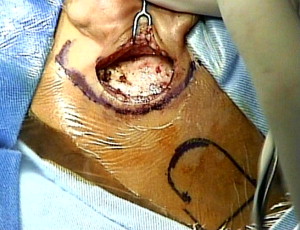
At Vanderbilt University and NYU, no effort is made to fix the electrode at the fantail or at the facial recess. Attempts are made to coil the redundant electrode array in the mastoid cavity, usually securing the coil in against the tegmen ( Fig. 3 ). A tight pocket technique for securing the R/S, snugged up with or without periosteal sutures, is currently preferred ( Fig. 4 ). This technique has shortened operative times, eliminating the need for (potentially biofilm-forming) additional foreign material. No significant R/S migrations have occurred at either center.
Minimally invasive techniques
Mastoidectomy with Posterior Tympanotomy Approach
In 1961, Dr House introduced the mastoidectomy with posterior tympanotomy approach (MPTA) for cochlear implantation. Since then, the MPTA has stood the test of time and become the most commonly used approach. As the name implies, a mastoidectomy is performed followed by a posterior tympanotomy, which opens the facial recess exposing the round window. Several techniques have been developed and explored to try to minimize the extent of surgery needed to place the implant and the risk to the facial nerve and chorda tympani associated with the MPTA.
Suprameatal Route
In 1999, Kronenberg and colleagues developed a technique that avoids a mastoidectomy altogether and introduces the electrode into the middle ear via a suprameatal route. This suprameatal approach is based on a retroauricular tympanotomy approach to the middle ear in which the facial nerve is protected by the body of the incus. Drawbacks to the suprameatal approach include the following :
- •
The electrode is stretched during insertion into the cochleostomy.
- •
Low-lying dura is a relative contraindication.
- •
A round window insertion and inferior cochleostomy is difficult.
- •
Additionally, the revision surgery rate is much higher with this technique (J. Thomas Roland Jr, MD, personal communication, 2011).
Endaural Approach
Another nonmastoidectomy technique uses an endaural approach for access to the cochleostomy and a superoposterior transcanal wall approach for the electrode. This endaural approach, also known as the Veria operation, requires a special perforator for drilling a direct tunnel and a safety electrode forceps for inserting the electrode.
Minimal Access Incision Techniques
Minimal access incision techniques have also been described. A percutaneous cochlear implant technique that involves a single, image-guided drill passed from the mastoid cortex through the facial recess to access the cochlea has been developed. The percutaneous cochlear implant technique uses an intraoperative CT scan and three fiducial markers in the bone surrounding the mastoid to plan a safe trajectory for the drill and has been validated in vitro and in vivo. Access to correct cochleostomy or round window insertion may also be limited and the 3-D approach to scala tympani insertion is limited.
Stay updated, free articles. Join our Telegram channel

Full access? Get Clinical Tree


