Purpose
To report the clinical features and surgical management of aniridic fibrosis syndrome using the type I Boston Keratoprosthesis (KPro).
Design
Interventional case series.
Methods
Retrospective chart review of 9 eyes in 9 patients with congenital aniridia that developed aniridic fibrosis syndrome.
Results
All patients had clinical diagnosis of congenital aniridia. Previously, all patients had undergone cataract surgery with posterior chamber intraocular lens (IOL) implantation and 7 patients had existing tube shunts. In all cases, fibrosis presented as progressive retrocorneal and retrolenticular membrane formation causing displacement of the IOL and secondary corneal decompensation. Two eyes had tractional folds in the retina with posterior extension of the membrane. The management included IOL explantation in 7 of 9 cases, removal of fibrosis with pars plana vitrectomy in all 9 patients, and implantation of a type I Boston KPro in all eyes. At a mean final follow-up of 26.1 months (range 6 to 48 months), vision remained improved in all patients. No patient had recurrence of the fibrotic membrane after KPro implantation.
Conclusion
This study represents another case series describing aniridic fibrosis syndrome and the largest study to report utilization of the type I Boston KPro in such patients. As the fibrosis can cause IOL dislocation, corneal decompensation, hypotony, and retinal detachment, monitoring for aniridic fibrosis syndrome in congenital aniridia with early surgical intervention is recommended. Type I Boston KPro may be considered in the surgical treatment of this condition.
Congenital aniridia is a panocular disease traditionally described as the classic triad of aniridia, foveal hypoplasia, and nystagmus. We now know that congenital aniridia is a spectrum of diseases resulting from mutations or deletions in PAX6 , the master control gene for eye development on chromosome 11p13. Congenital aniridia affects a variety of ocular structures including the cornea, anterior chamber, iris, lens, macula, and optic nerve. While foveal hypoplasia remains the major factor limiting visual acuity, many experience decline in visual acuity from the development of a presenile cataract (50%–85%), glaucoma (50%–75%), and aniridic keratopathy (20%).
Management of congenital aniridia usually begins medically; however, eventually all patients will need several surgical interventions to preserve or restore their baseline visual acuity. Implantation of intraocular lens (IOL) and tube shunts to control glaucoma are common. In addition, many will require limbal stem cell and corneal transplantation for aniridic keratopathy.
The corneal changes associated with aniridic keratopathy include vascular pannus formation, conjunctivalization, epithelial erosions, and subepithelial fibrosis. These corneal changes are primarily attributable to progressive limbal stem cell deficiency. Ocular surface reconstruction with limbal stem cell transplantation, either with or without corneal transplantation, provide long-term favorable outcomes in aniridic keratopathy by restoring limbal stem cell function. The use of the type I Boston Keratoprosthesis (Boston KPro) is a novel approach to manage aniridic keratopathy with favorable outcomes in terms of visual acuity and device retention. Additionally, keratoprosthesis eliminates the need for systemic immunosuppressive therapy required after limbal stem cell allograft.
Tsai and associates were the first to describe a postsurgical anterior chamber progressive fibrosis that occurred in patients with congenital aniridia who had undergone multiple intraocular surgeries. They termed the disease aniridic fibrosis syndrome. Our study represents a case series on the syndrome and describes the clinical features and surgical outcomes in 9 patients. In addition, we report the use of type I Boston KPro as a surgical option in this subset of aniridic patients.
Patients and Methods
A retrospective chart review of 4 eyes in 4 consecutive patients with congenital aniridia who developed aniridic fibrosis syndrome from March 2006 to December 2009 at the University of Illinois Eye and Ear Infirmary and 5 eyes in 5 patients at the Cincinnati Eye Institute from July 1999 to January 2010 were performed. Pertinent recorded data included age, sex, past ocular history including medical and surgical interventions, Snellen visual acuity, and clinical examination findings.
Surgical intervention included IOL explantation (7 cases) and removal of fibrosis with anterior vitrectomy (1 case) or pars plana vitrectomy (8 cases). In 7 of 9 eyes, a type I Boston KPro was implanted primarily. One eye had a Boston KPro prior to the development of fibrosis development, which was later repeated because of a corneal melt. In the remaining case, an Alphacor KPro (Argus Biomedical Pty Ltd, Perth, Australia) was exchanged for Boston KPro. Boston KPro (Massachusetts Eye and Ear Infirmary, Boston, Massachusetts, USA) was used in all cases with polymethyl metacrylate backplate of 8.5 mm in diameter.
Results
Patient data and outcomes are presented in the Table .
| Patient | Age/Sex/Eye | Previous Ocular Surgery | BCVA Before AFS | BCVA After AFS | Interventions After Diagnosis of AFS | BCVA After KPro | Recurrence-Free Interval After KPro (months) |
|---|---|---|---|---|---|---|---|
| 1 | 11/F/OS | Tube shunts OU, PCIOL OS | 20/400 | CF 3 ft | IOL explantation/anterior vitrectomy/membrane removal/Boston KPro | 20/400 | 6 |
| 2 | 10/M/OS | Tube shunts OU, PK OS ×2, PCIOL OS sutured | 20/400 | HM | IOL explantation/PPV/Boston KPro | 20/400 | 36 |
| 3 | 20/F/OS | Tube shunt OS ×2, RD repair OS, PCIOL OS, PK OS, KLAL OS, DSAEK OS, Boston KPro OS | 20/400 | HM | IOL explantation, PPV/Boston KPro | 20/400 | 12 |
| 4 | 67/M/OD | PCIOL, AM for corneal melt, KLAL | HM | HM | IOL explantation/PPV/tube shunt/Boston KPro | 20/200 | 12 |
| 5 | 11/F/OD | PK, Phaco/IOL, KLAL, PK ×2, tube shunt | 20/200 | CF | PK/ant synechiolysis, PPV/membrane peel/IOL removal/revision of tube shunt, KLAL, Boston KPro | 20/500 | 48 |
| 6 | 71/M/OD | Cataract extraction, Morcher IOL, KLAL, tube shunt, PK ×2, tube revision ×2 | 20/160 | HM | Boston KPro, PPV/ removal of retroprosthetic membrane, PPV/repair of tractional RD/removal of cyclitic membrane encasing Morcher IOL/tube revision/IOL repositioning | 20/400 | 27 |
| 7 | 41/F/OD | KLAL ×2, tube shunt, PK/Phaco/Morcher IOL | 20/60 | CF | PK/IOL exchange/membrane removal, PK/PPV/membrane removal, PK/PPV/IOL explantation/membrane removal, Boston KPro, YAG fibrotic membrane, PPV/cyclitic membrane removal/tube ligation | 20/250 | 25 |
| 8 | 60/F/OD | KLAL ×2, PK ×2, tube shunt, Phaco/IOL, Alphacor KPro, Alphacor KPro explanted/PK | 20/250 | CF 6 ft | Boston KPro with IOL explantation, YAG fibrotic membrane, Boston KPro exchange/PPV/membrane peel | CF 2 ft | 40 |
| 9 | 61/F/OS | KLAL, PK, Phaco/IOL | 20/200 | CF | Boston KPro/IOL explantation, YAG retroprosthetic membrane, PPV/removal cyclitic membrane | 20/250 | 29 |
Representative Case Report (Case 1)
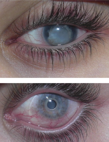
An 11-year-old female patient with congenital aniridia was referred for a left corneal opacity and gradual decline in visual acuity over 3 months. Ocular history was significant for secondary glaucoma in both eyes with tube shunts placed 6 and 2 years ago in the right and left eyes, respectively. Family history was positive for aniridia. Visual acuity in the left eye was count fingers at 3 feet, which had declined from 20/400. Intraocular pressure was 10 mm Hg. The cornea was cloudy and edematous. Anterior and posterior chamber examination revealed a dense retrocorneal and retrolental membrane with anterior displacement of the posterior chamber intraocular lens (PCIOL). Posterior examination was limited by corneal opacity and the anterior segment membrane. A diagnosis of aniridic fibrosis syndrome was made and the patient underwent IOL explantation and anterior vitrectomy. Postoperatively, visual acuity improved to 20/400. However, at 12 months the membrane recurred and visual acuity decreased to hand motions. On examination, the patient had significant corneal decompensation and anterior chamber fibrosis ( Figure , Top). The patient then underwent pars plana vitrectomy, membrane removal, and implantation of a Boston KPro. At 6 months after the KPro surgery, a thin retrosprosthetic membrane was observed and successfully cleared by yttrium-aluminum-garnet laser, improving her visual acuity to 20/200 ( Figure , Bottom).
Summary of Findings
All 9 patients were diagnosed with congenital aniridia based on clinical features. One patient had WAGR syndrome. The patients ranged in age from 11 to 71 years (mean 33.8 years). All patients had previous cataract surgery with PCIOL implantation. Seven of the 9 patients had existing tube shunts for glaucoma. Seven patients had previous corneal transplantation, 7 had previous keratolimbal allograft, 1 had an existing Boston KPro, and 1 had existing Alphacor KPro.
In all eyes, fibrosis presented as progressive retrocorneal and retrolenticular membrane formation causing anterior displacement of the intraocular lens and endothelial cell damage with corneal decompensation. One patient experienced a corneal melt and perforation with contracting fibrous tissue anteriorly displacing the IOL into the cornea. Two patients had tractional retinal folds attributable to posterior extension of the membrane. The initial management in all patients (except Patients 3 and 6) included IOL explantation and removal of fibrosis with anterior vitrectomy (1 patient) or pars plana vitrectomy (8 patients). In 7 of 9 cases, a type I Boston KPro was primarily implanted. The patient with the anterior vitrectomy did not initially receive a KPro, but later experienced progressive corneal decompensation after IOL explantation and subsequently underwent complete pars plana vitrectomy with KPro implantation. In Patient 6, the Morcher lens was too severely fibrosed to the intraocular structures for safe removal. One patient (Patient 3) developed fibrosis in the setting of a previous KPro and later underwent repeat KPro implantation because of melting of the carrier cornea tissue. In another eye, an Alphacor KPro, which was initially explanted for a penetrating keratoplasty, was eventually replaced with a Boston KPro (Patient 8). In 7 patients an aphakic KPro was used while in 2 cases the IOL was retained and a pseudophakic KPro was used. All devices (100%) were retained throughout the entire follow-up period of 6 to 48 months (mean 26.1 months).
After KPro implantation, vision improved in all eyes, ranging from hand motions to count fingers preoperatively, to 20/200 to 2/500 postoperatively. One patient (Patient 4) was stable after KPro implantation for 12 months with vision of 20/200, but after silicone oil removal, the patient developed a suprachoroidal hemorrhage and eventually vision became no light perception. In Patient 6, where the fibrosed Morcher IOL was retained after KPro implantation, both tractional retinal detachment and IOL dislocation occurred, which required endoscopic repair. Postoperative events after KPro implantation included retroprosthetic membrane in 5 eyes (Patients 1, 6, 7, 8, and 9), suprachoroidal hemorrhage in 1 eye (Patient 4), and tractional retinal detachment in 1 eye (Patient 6). The Figure shows a preoperative and postoperative photograph of Patient 1.
Stay updated, free articles. Join our Telegram channel

Full access? Get Clinical Tree


