I was interested to read the study by Xu and associates. They concluded that in age-related macular degeneration (AMD), “development and progression are associated with decreased choroidal circulatory parameters.” They go on to state: “All these findings suggest a possible role for choroidal circulatory abnormalities in the development of AMD and CNV [choroidal neovascularization].”
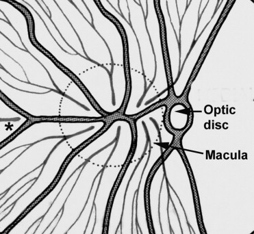
Almost 40 years ago, I investigated, by fluorescein fundus angiography, the circulation of the submacular choroidal vascular bed in vivo. I discovered that there are watershed zones between the regions supplied by the various short posterior ciliary arteries and that all the watershed zones of the temporal short posterior ciliary arteries meet in the submacular choroid ( Figure ). The significance of the watershed zones is that in the event of a fall in the perfusion pressure in the vascular bed, the watershed zone, being an area of comparatively poor vascularity, is most vulnerable to ischemia. Watershed zones are known to play an important role in ischemic disorders, for example, ischemic infarcts in the brain and anterior ischemic optic neuropathy. Based on my findings about the location of the watershed zones in the submacular choroid, I then stated: “The present studies, therefore, suggest that the macular region is especially vulnerable to chronic ischaemic disorders.” I further stated: “In the light of my findings the frequent occurrence of senile macular degeneration (now AMD) is not at all surprising. In senile macular degeneration, the well documented submacular choroidal neovascularization from the choroidal vascular bed may represent a response to chronic ischaemia.” This concept immediately was discredited and long forgotten. (Xu and associates did not mention it. ) Furthermore, in fluorescein angiography in early AMD, I reported a filling delay of the watershed zones between the temporal short posterior ciliary arteries in the center of the macular region. There is other evidence suggesting that submacular choroid is more vulnerable to ischemic disorder than other parts of the choroid; for instance, in hypertensive choroidopathy and on experimental reduction of perfusion pressure in the choroid vascular bed, fluorescein angiography showed delayed or no filling of the watershed zones in the submacular choroid and of the choriocapillaris in that region. Ross and associates, examining indocyanine green angiography, showed an increased incidence of choroidal watershed zone filling defects in patients with AMD and postulated that that may predispose them to choroidal neovascularization. A reduction in perfusion pressure and blood flow in a tissue can be influenced by several systemic and local factors. In the case of AMD, that further adds to its multifactorial etiology.