Salivary gland preservation during treatment for obstructive duct and gland problems is a goal worth pursuing. Difficult cases may seem to be candidates for sialadenectomy. However, progress in endoscopic and open-surgical procedures can help the physician to find solutions that overcome difficult problems without removing the gland. Broader application of these sialendoscopic and open preservation procedures may be especially useful for physicians without access to extracorporeal lithotriptors.
Salivary gland surgery has progressed to the point where many pathologies that once required gland resection are now removed worldwide with endoscopes and extracorporeal lithotriptors. Nevertheless, patients can present to their physician with extraordinary salivary duct pathologies. The diagnosis and treatment for these difficult cases can lie outside the usual clinical treatment algorithms for endoscopes and lithotriptors. At first, the best treatment for these difficult problems might seem to be surgical gland resection. However, the goal of achieving physiologically intact salivary glands after treatment runs contrary to gland removal. The physician is placed in the challenging position of finding ways to overcome salivary duct problems without removing the gland. As an additional complicating factor, in North America, extracorporeal shockwave lithotripsy (ESWL) for the salivary gland is not yet governmentally approved; the published algorithms for physicians and patients from other countries depart from the North American experience. Therefore, some alternative treatment options have been developed for difficult cases of salivary duct obstruction in North America, of which several are applicable to patients anywhere.
Endoscopic segmental-open approach
Some patients with larger stones cannot be treated effectively solely by intraductal approaches. Nahlieli, in 2002, and McGurk in 2004, described parotid procedures combining endoscopic intraductal transillumination of the skin, as a surgical target locator, with an open-surgical approach, to gain direct access to the stone and deliver it from the duct. After removal of the stone, the duct is closed over a stent. These 2 approaches work well for Stensen duct or hilum stones, but they do not address the “intraparenchymal” stone located in the secondary and tertiary duct or the diseased salivary drainage basin proximal to the stone. In the author’s institution, experience has shown that the so-called intraparenchymal stones are probably intraductal, with the actual duct being expanded until transparently thin and the stone appearing to be in the parenchyma. In these proximal areas, no stent can be placed for postoperative conservation of the duct. In addition, the wedge-shaped drainage-basin of the gland proximal to these stones is often already chronically diseased with severe ductal ectasia. Conversely, the uninvolved ducts and their attached gland tissue may be normal. Therefore, a parotidectomy would needlessly remove normal tissue along with diseased gland. For cases of a stone within an isolated duct that blocks a diseased drainage basin, Endoscopic Segmental-Open parotidectomy is introduced.
Segmental Parotidectomy
The patient is prepared for surgery as per McGurk, with the addition of a nerve integrity monitoring system (NIMS; NIM-Response 2.0, Medtronic, Inc, Jacksonville, FL, USA) facial nerve monitor. The stone is endoscopically located where it is lodged in the specific secondary or tertiary duct. Through the gland tissue, the stone is dissected free of the duct and parenchyma and removed. Thereafter, the remaining diseased duct and drainage basin are resected by performing a wedge-shaped partial parotidectomy. The partial parotidectomy targets only the diseased gland segment. The other normal ducts and gland tissue are left intact. By removing the stone and the specifically involved ducts and gland tissue, a repetitive cycle of sialadenitis is avoided.
Regarding cosmesis, the real effect of a parotidectomy is seen when resected areas cause a facial depression and create areas of shadow. Most noticeable is the “eye-catcher” shadow located below the zygomatic arch. The parotid gland in the zygomatic area is rarely found to have stone pathology or other problems; its duct and parenchyma can usually be spared to great cosmetic advantage for the patient. The residual normal gland effaces the light shadow and leads to markedly improved cosmesis.
Sometimes, there may be areas of zygomatic parenchyma that have no duct connections but are left for cosmetic reasons. If the isolated gland tissue is bulky, it can be injected with botulinum toxin type A to render the tissue inactive and prevent sialocele, until the raw tissue bed heals and scar tissue obliterates the surgical planes.
Another advantage of the segmental parotidectomy using an endoscope is that the possibility of a sialocele is greatly reduced by meticulous closure of the circumscribed surgical field. Any open ducts and raw gland tissue are given attention. Because the endoscope is already positioned within the duct, after the resection of parotid tissue is completed, back-pressure testing of the duct with saline solution is performed. It allows the surgeon to find and oversew even the smallest ducts that were crosscut in the dissection. To help protect against Frey syndrome, a pie-shaped wedge of a collagen matrix barrier material (Duragen, Integra Lifesciences Corporation, Plainsboro, NJ, USA) is sewn into the corresponding wedge of parotid bed.
Patient 1
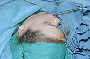
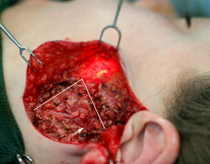
An 11-year-old female patient, with recurrent left parotid swelling for 4.5 years, underwent sialography showing an 11.6 by 8.0 mm stone in a secondary duct. Sialendoscopy could visualize the stone, but not reach it for intraductal instrumentation treatment. No ESWL lithotriptor was available. A Transfacial approach through the cheek was not allowed by the family for cosmetic reasons, which would also not have been able to address the proximal gland drainage basin and facial nerve. Because the stone was in a secondary duct, the possibility of duct stenoses, with future surgery needed to treat residual gland problems, was a major consideration. The Endoscopic-Open approach, with endoscopic transillumination and NIMS facial nerve monitoring, was used to find and remove the stone through a modified-Blair incision ( Fig. 1 ). The stone “socket” with its specific salivary drainage basin was then segmentally removed ( Fig. 2 ). The unaffected upper half of the superficial lobe was preserved. The remaining crosscut ducts were back-pressured with saline solution and oversewn with 5-0 chromic sutures. The remaining upper and lower parts of the anterior gland parenchyma were loosely closed in a “V to Y” fashion. A collagen barrier was placed. The remaining zygomatic area parotid gland provided sufficient tissue to offset a cosmetic parotid defect; a smooth transition between the zygoma and the cheek contours was achieved ( Fig. 3 ). Postoperatively, the parotid orifice showed clean saliva when “milking” the remaining gland. A 5-year follow-up showed the patient to be asymptomatic, with a functioning residual gland on physical examination.

Endoscopic double-open approach
At times, the anteriorly positioned Stensen duct and the posterior-most parotid gland may be simultaneously diseased. In these difficult cases, anterior duct structures may need to be surgically accessed, in combination with surgical work on the posterior gland and the facial nerve. Therefore, access to the entire length of the parotid duct and gland is needed.
The classic preauricular parotidectomy incision, with undermining in an anterior direction, does not allow for dissection of the anterior parotid duct and facial nerve branches. An increasingly narrow surgical field is found in the anterior acute angle between parotid gland and skin. Visualization of the anterior nerve and duct is poor. The main facial nerve branch that overlays the Stensen duct is the buccal branch; however, other nerve arborizations can be prolific and only direct transfacial access provides a surgical field for reliable nerve preservation. Conversely, if a transfacial incision is made at a midmasseter muscle location, then dissection over the posterior parotid gland and facial nerve is not possible; a preauricular approach is also needed.
To achieve effective surgical exposure over the entire parotid gland and duct, the “Endoscopic Double-Open” approach is introduced. The transfacial and modified-Blair incisions are used during the same procedure. The transfacial incision allows the facial nerve main buccal branch, the cobweblike facial nerve arborizations, and the anterior duct to be readily visualized and surgically operated. The preauricular incision allows surgical access to the posterior portion of the parotid gland and facial nerve that are beyond the posterior limits of the transfacial incision. With the endoscope in the duct and the 2 external incisions, the surgeon can feel confident of having complete surgical exposure from inside and outside, and from the anterior duct to the posterior duct and gland. This technique gives the surgeon complete access to the parotid duct and gland.
Patient 2
A 24-year-old man presented with a progressive 3-year history of a 6.0 by 3.0 cm subcutaneous tubular mass of the right cheek. A magnetic resonance scan showed the appearance of a giant sialocele ( Fig. 4 ). The gland itself had ectasias. The other major salivary glands were normal. Using the endoscopic technique, the duct showed a complete, nondistensible, “hard” stenosis at the anterior border of the masseter muscle. The holmium laser was used to penetrate the stenosis and the sialocele was entered. Much turbid, green-colored, salivary mucus with thickly particulate sediment was encountered inside the sialocele ( Fig. 5 ). The walls became heavily rugated as the sialocele was decompressed. Large sedimentary flakes of green matter detached from the walls. The proximal end of the sialocele led into the hilum and stenotic duct branches. It was clear that a hollow stent would become recurrently plugged. Also, an intraoral sialocelostomy would probably be prone to retrograde infection because of the cavernous size of the sialocele and poor condition of the gland. A vein graft replacement appeared to have a poor chance of success, because of the dilated hilum deep within the gland and multiple intraglandular duct stenoses. Also, the patient wanted a “cure” at one surgical sitting, rather than recurrent attempts to rehabilitate a chronically diseased gland. Rather than continue a chronic parotid problem, the decision was made to segmentally resect the gland and the sialocele duct using the Endoscopic Double-Open approach. The endoscope was used to transilluminate the decompressed sialocele and the skin, and help mark the course of the duct and locate the Transfacial incision. The NIMS monitor was in place and a microscope was used to carefully transfacially dissect the anterior-most facial nerve branches. The duct was dissected forward into the mouth and freed up to the parotid hilum posteriorly ( Fig. 6 ). Through the preauricular incision, a Segmental parotidectomy with facial nerve preservation was performed; the segment of gland tissue abutting the zygoma was left in place to atrophy and help the cosmetic effacement of the defect. A collagen barrier was placed over the raw tissue bed to help prevent Frey syndrome. The incisions were closed using plastic technique ( Fig. 7 ). The patient is asymptomatic 2 years after surgery, without facial nerve or cosmetic deficits.
Stay updated, free articles. Join our Telegram channel

Full access? Get Clinical Tree


