(1)
St. Johns, FL, USA
(2)
Helen Keller Foundation for Research and Education, International Society of Ocular Trauma, Birmingham, AL, USA
(3)
Consultant and Vitreoretinal Surgeon, Milos Eye Hospital, Belgrade, Serbia
(4)
Consultant and Vitreoretinal Surgeon, Zagórskiego Eye Hospital, Cracow, Poland
In principle, it would be preferable not to employ scleral indentation since it distorts the anatomy and the surgeon is forced to perform his maneuvers in an artificially altered environment. In reality, the only way to avoid scleral indentation as the surgeon works in the periphery is to use the endoscope for viewing (EAV, see Sect. 17.3). In traditional PPV, however, scleral indentation is a necessity if the surgeon needs to access the anterior part of the vitreous cavity.
28.1 The Advantages of Scleral Indentation
Normal structures, which would remain hidden from view otherwise, become accessible and their pathologies treatable. The structures include the peripheral retina, the vitreous base, and the ciliary body.
A bullous or highly mobile retina, if indented, will have reduced mobility. The safety margin of PPV increases both on the “hilltop” and on the “slopes” (see below and Fig. 28.1).
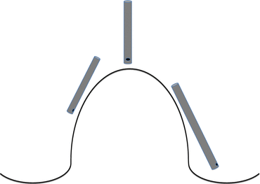
Fig. 28.1
Cross-sectional view of performing vitrectomy during scleral indentation. The internal elevation created by the indentation of the sclera is not cone-shaped. The effect is a radially oriented “mountain” or a circumferentially oriented “hill” with typically symmetric slopes on either side. The more peripheral the indentation, the less radial the direction of the ridge. If the retina was attached before, it will remain so during the indentation (shown here). If the retina was detached before and the detachment is not too high, the indentation results in a (temporary) retinal reattachment on the hilltop, but the RD persists on the slopes. Vitrectomy should be performed not only over the hilltop but also on the two slopes. The angle of the probe’s shaft relative to the retina changes according to the location the indentation and the site of the sclerotomy (see the text for more details). The surgeon must decide, based on the visual feedback of his actions, whether to turn the port toward, parallel with, or away from the retina. He must also change the settings of the vitrectomy machine according to tissue behavior
28.2 The Mechanics of Vitrectomy with Scleral Indentation
The contour of the eyeball dramatically changes with scleral indentation. If the retina was attached, what had a concave profile before becomes a convex one; if the retina was detached, it is now reunited with its foundation.
The angle between the probe’s shaft and the retina changes; consequently the port’s plane relative to the retinal surface also changes. The person performing the indentation must understand its mechanics as well as the risks involved (see below and Sect. 24.1). Retinal injury can result in multiple scenarios.
Lack of coordination between the external vs the internal movements1 (see below).
Unexpected changes in the characteristics of the indentation while the intraocular tissues are being manipulated are very dangerous.
Pearl
Four variables should be taken into account to accurately describe the characteristics of the indentation: height (depression of the eyewall into the vitreous), location (according to clock hours in the frontal plane), depth (how far posterior from the ora serrata), and angle (this is the variable that changes the least; typically, the direction is radial).
A sudden increase in the height or location of the indentation.2 A common cause of slippage is a lid speculum with a long blade (see Fig. 19.3). When the area of contact between speculum and lid is small, the depressor can be inserted on either side of it and there is no slippage. When moving the depressor along the long blade in the darkness, however, the nurse has no feedback that she reached the blade’s edge until it is too late and the depressor suddenly “jumps.”
A surgeon too focused on the port of the probe. If the vitrectomy is done in a central location, the shaft may rub against the peripheral retina and cause an erosion (break) or bleeding.
Incorrectly set PPV parameters. The aspiration/flow is too high, not adjusted to the peculiar demands of the periphery (see Table 12.2); a retinal break or detachment may result.
A surgeon who does not take into consideration the implications of the change in the eyewall’s contour (see below).
In a pseudophakic/aphakic eye, the surgeon must decide whether to switch hands to complete the process. Table 28.1 shows the consequences if the probe remains in the same sclerotomy.
Table 28.1


The implications for the execution of peripheral vitrectomy according to the location of the scleral indentation*
Stay updated, free articles. Join our Telegram channel
Full access? Get Clinical Tree
