Purpose
To investigate the relationship between systemic cytokines, the complement factor H ( CFH ) Y402H polymorphism, drusen load, and subfoveal choroidal thickness in patients with dry age-related macular degeneration (AMD).
Design
Cross-sectional study.
Methods
Forty-four dry AMD patients under care of the Retina Service at the University of British Columbia were enrolled. Drusen load was measured with an automated software algorithm in spectral-domain optical coherence tomography; subfoveal choroidal thickness was measured manually using enhanced depth imaging. Bio-Plex suspension assays (Bio-Rad Laboratories) were used to analyze cytokines in plasma and CFH Y402H was genotyped. Statistical analyses included analysis of covariance and Pearson correlation, corrected for multiple comparisons.
Results
The levels of 3 of 4 studied cytokines were significantly different among patients with CC, CT, or TT variants of the CFH Y402H polymorphism ( P < .01). Patients with the at-risk CC variant had higher systemic levels of interleukin-6, interleukin-18, and tumor necrosis factor α than those with the CT variants, the TT variant, or both ( P < .01). Interleukin-1β did not reach significance ( P = .02), but did demonstrate a consistent trend. No correlation was found between plasma cytokines and drusen load or choroidal thickness (all P > .15).
Conclusions
The elevated systemic levels of selected proinflammatory cytokines, including those representing products of inflammasome activation, were associated with the CC at-risk variant of the Y402H polymorphism and suggest that genetic factors regulate the inflammatory status in dry AMD patients. Our data support the central role of inflammation in the pathogenesis of AMD and provide further evidence of a systemic involvement in AMD etiology.
Age-related macular degeneration (AMD) constitutes the number 1 cause of blindness among the elderly in industrialized countries. The hallmark of AMD is the accumulation of extracellular deposits called drusen, which are located between the retinal pigmented epithelium (RPE) and Bruch membrane. Larger size and greater number of drusen confer higher risk of developing vision-loss due to geographic atrophy and choroidal neovascularization. In addition, choroidal abnormalities have been reported to be associated with drusen formation in dry AMD. The recent introduction of enhanced depth imaging and segmentation algorithm in spectral-domain optical coherence tomography (SD-OCT) allows for better quantification of drusen load and choroidal measurements. This noninvasive, reliable imaging method of measuring drusen load and choroidal thickness creates an opportunity to quantify these parameters of slow progression in patients with the early stage of dry AMD. Additionaly, this method has the potential to allow for an in-depth study of the underlying mechanisms in AMD pathogenesis.
Although the clinical presentation of AMD mainly involves local processes within the retina, systemic factors in the circulating blood also may contribute to the pathogenesis of AMD via exchange between the choroid and the retina. For example, the homozygous CC variant of the Y402H polymorphism in the gene coding complement factor H ( CFH ), a regulator of the alternative complement pathway, is associated with a high incidence as well as progression to late AMD. Proposed mechanisms supporting this include the finding that circulating CFH at-risk proteins have a weaker capacity to bind oxydized phospholipids in situ, thereby modulating proinflammatory stress in the outer retina. Systemic activation of the complement cascade also was found in AMD patients. Together with the crosstalk between the complement activation products and proinflammatory cytokines reported in blood cells, these findings highlight the potential role of systemic inflammatory cytokines in the pathogenesis of AMD. However, several questions remain because few studies have addressed the relationship between the systemic cytokine levels and the CFH Y402H polymorphism in AMD patients, or whether systemic levels of cytokines are associated with the local ocular manifestations such as drusen load and choroidal thinning that occur in early dry AMD. Using a new SD-OCT tool to follow drusen enlargement and changes of choroidal thickness, 2 parameters used to assess disease progression, we conducted a pilot study to investigate the relationship of systemic cytokines with these 2 parameters as well as the CFH Y402H polymorphism in patients with dry AMD. We focused on 4 inflammation-related cytokines. Interleukin (IL)-1β, IL-6, IL-18, and tumor necrosis factor α (TNF-α) are classic proinflammatory cytokines. Previously, they were shown to affect RPE function and were implicated in AMD pathogenesis.
Methods
Study Population
The design of this study was cross-sectional, and it was approved prospectively by Providence Health Care Research Ethics Board at the University of British Columbia and complied with the tenets of the Declaration of Helsinki. Forty-four patients under the care of the physicians at the Retina Service at University of British Columbia (D.A.A., F.F., A.K., and A.B.M.) were enrolled in this study with informed consent. The inclusion criteria were age 55 years or older, diagnosis of dry AMD in both eyes, and absence of any other disease affecting the macula that could compromise the ability to assess properly for drusen (eg, epiretinal membrane, vitreomacular traction, adult vitelliform dystrophy, etc). Exclusion criteria were exudative AMD or geographic atrophy in either eye; ocular media opacity that precluded adequate visualization or imaging of the macula; cancer within the previous 10 years (except for basal cell carcinoma); cardiovascular disease with complications (heart attack or stroke) within the previous year; diabetes with uncontrolled glucose level or complications (diabetic eye disease, diabetic kidney disease, or diabetic nerve damage); any other medical conditions that, in the opinion of the investigators, would affect the study results; and medications that could affect the levels of biomarkers in blood (eg, corticosteroids and NSAIDs).
Eye Examination, Drusen Area, and Choroidal Thickness Measurements
Patients underwent a full ophthalmic examination including measurement of best-corrected visual acuity, intraocular pressure, biomicroscopy, and dilated fundus examination. Cirrus SD-OCT (version 6 software; Carl Zeiss Meditec, Inc., Dublin, California, USA) was used for drusen area and choroidal thickness measurements. Each eye was scanned once using a cube scan protocol (200 × 200 cube scan). In the same session, each eye also was scanned a minimum of twice with the enhanced depth imaging raster scan. Each scan was centered on the fovea automatically and covered a 6 × 6-mm area. The minimum required signal strength for every scan was 6 of 10.
The drusen areal measurements were generated automatically by the Cirrus SD-OCT software algorithm from the cube scans. For measurement of subfoveal choroidal thickness, 3 independent investigators (A.K., K.P.-V., F.F.) measured the choroidal thickness beneath the fovea manually from the enhanced depth imaging raster images using the program’s caliper function during 2 different sessions. The scan with the best signal strength score was chosen for each eye. Each researcher was blinded to the measurements of others and to their own previous sessions.
Cytokine Analysis
Nonfasting blood specimens were obtained at the time of the study visit. Ethylene-diamine-tetraacetic acid plasma from patients was prepared into aliquots within 2 hours of phlebotomy and was stored at −80°C until analysis. Cytokines of ethylene-diamine-tetraacetic acid plasma from patients were measured by Bio-Plex Pro human cytokine, chemokine, and growth factor assays as described by the manufacturer (Bio-Rad Laboratories, Hercules, California, USA). The assays use xMAP technology (Luminex, Austin, Texas, USA), which permits the quantification of multiple cytokines in a single well with 50 μL of diluted sample (1:4). In our experiments, the premixed multiplex beads of the Bio-Plex human cytokine 27-plex assay and an additional 2-plex were used. They included the following cytokines: Interleukin (IL)-1β, IL-1ra, IL-2, IL-4, IL-5, IL-6, IL-7, IL-8, IL-9, IL-10, IL-12 (p70), IL-13, IL-15, IL-17, IL-18, chemokine (C-C motif) ligand (CCL)11/eotaxin, basic fibroblast growth factor (basic FGF), granulocyte colony-stimulating factor (G-CSF), granulocyte macrophage colony-stimulating factor (GM-CSF), interferon-gamma (IFN-γ), chemokine (C-X-C motif) ligand (CXCL)10/interferon gamma-induced protein 10 (IP-10), CCL2/monocyte chemoattractant protein (MCP)-1, CCL3/macrophage inflammatory protein (MIP)-1α, CCL4/MIP-1β, platelet-derived growth factor (PDGF)-BB, CCL5/regulated on activation, normal T cell expressed and secreted (RANTES), CXCL12/stromal cell-derived factor (SDF)-1α, tumor necrosis factor (TNF)-α, and vascular endothelial growth factor (VEGF).
Complement Factor H Y402H Genotyping
Genomic DNA was extracted from ethylene-diamine-tetraacetic acid-contained whole blood using QIAsymphony SP system (Qiagen, Toronto, Canada) and was eluted in 200 μL volume. The extracted DNA was quantified using the Quant-iT PicoGreen dsDNA Assay Kit (Life Technologies, Burlington, Canada) to make sure that a sufficient concentration (> 50ng/μL) of genomic DNA was obtained. The subsequent polymerase chain reaction (PCR) amplification and sequencing analysis were performed as previously described. Briefly, amplicons of 660 bp containing the CFH Y402H polymorphism (rs1061170) were produced in PCR reaction using the forward and reverse primers of 5′ AGTAACTTTAGTTCGTCTTCAG 3′ and 5′ ATCTTCTTGGTGTGAGATAACG 3′, respectively. After being purified with the QIAquick PCR Purification Kit (Qiagen), the PCR products were mixed with sequencing primer at suitable concentrations as required and then sent to GENEWIZ (South Plainfield, New Jersey, USA) for sequencing analysis. The CFH sequencing primer was 5′ ACTTTAGTTCGTCTTCAG 3′.
Statistical Analysis
Means were calculated between the 2 eyes for drusen area, as well as between the 2 eyes and the 3 measurements for choroidal thickness. We used a mean value to compare with the single measurements obtained from the cytokines. All analyses were conducted with SPSS Statistics version 20 (IBM, Armonk, New York, USA). Our previous study showed that SD OCT-based drusen area and drusen volume, 2 parameters of drusen, are highly correlated ( r = 0.86). For this reason, we decided to use only drusen area in our analysis.
In the first analysis, we examined the differences in plasma levels of 4 cytokines among the 3 variants of the CFH Y402H polymorphism in our study population. We conducted a series of analyses of covariance using the genotype groups (CC, CT, TT) as the between-subjects factor and individual cytokine, drusen area, and choroidal thickness as the dependent variables. We also included age as a covariate because previous work showed the possible relationship between cytokines and age. We used a Bonferroni correction to account for multiple comparisons ( P < .01) and post hoc comparisons were conducted after a significant omnibus result. For the post hoc comparisons, we used a priori contrasts between the CC and CT variants and between the CC and TT variants, because we were most interested in these contrasts.
In the second analysis, we examined the correlation between each selected cytokine and drusen area or choroidal thickness. We conducted a series of Pearson correlations using cytokines, drusen area, and choroidal thickness as variables. We again used a Bonferroni correction to account for multiple comparisons ( P < .01).
Results
Subject Characteristics
Participants for this study were recruited between February 2012 and May 2012. The characteristics of the 44 subjects were described previously. Briefly, each subject had bilateral dry AMD. None of our subjects were current smokers. The mean age of all subjects was 75.77 years (standard deviation, 8.42 years). The mean drusen area between the 2 eyes was 1.44 mm 2 (standard deviation, 1.22 mm 2 ), with 1.57 mm 2 (standard deviation, 1.64 mm 2 ) for right eyes and 1.30 mm 2 (standard deviation, 1.16 mm 2 ) for left eyes. The mean choroidal thickness between the 2 eyes was 230.73 μm (standard deviation, 53.23 μm), with 232.00 μm (standard deviation, 53.44 μm) for right eyes and 229.47 μm (standard deviation, 59.58 μm) for left eyes. In terms of the CFH Y402H polymorphism, 17 patients (38.6%) had the CC variant, 14 patients (31.8%) had the CT variant, and 13 patients (29.5%) had the TT variant ( Table 1 ).
| Sample Characteristics | Mean (SD) or No. (%) | 95% CI |
|---|---|---|
| Age (y) | 75.77 (8.42) | 73.28 to 78.26 |
| Drusen area (mm 2 ) | 1.44 (1.22) | 1.08 to 1.80 |
| Right eyes | 1.57 (1.64) | 1.09 to 2.05 |
| Left eyes | 1.30 (1.16) | 0.96 to 1.65 |
| Choroidal thickness (μm) | 230.73 (53.23) | 215.00 to 246.46 |
| Right eyes | 232.00 (53.44) | 216.21 to 247.79 |
| Left eyes | 229.47 (59.58) | 211.86 to 247.07 |
| CFH Y402H genotype a | ||
| CC | 17 (38.6%) | |
| CT | 14 (31.8%) | |
| TT | 13 (29.5%) |
a CC, CT, and TT are 3 genotype variants of CFH Y402H polymorphism.
Cytokines, Drusen Area, Choroidal Thickness, and Genotype Variants
To examine if there is a relationship between systemic cytokines, drusen area, or choroidal thickness and genotype of the CFH Y402H polymorphism, 4 cytokines were selected for analysis: IL-1β, IL-6, IL-18, and TNF-α. IL-1β and IL-18 were both products of inflammasome activation, a pathway recently studied for its association with both geographic atrophy and choroidal neovascularization development. IL-18 was known to induce RPE degeneration in mice. IL-6 and TNF-α are classic proinflammatory cytokines that have been shown to affect RPE function in vitro.
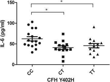
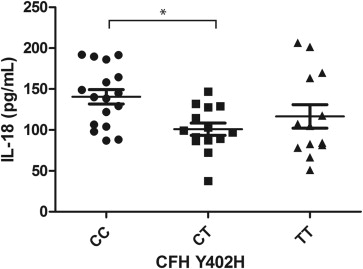
Our analysis of covariance results showed that 3 of the 4 cytokines were significantly different among patients with the CC, CT, or TT variants of the CFH Y402H genotype ( P < .01), when corrected for age ( Table 2 ). Patients with the CC variant had a higher level of IL-6 than those with the CT or TT variant ( P < .01). The level of IL-6 in patients with the CC variant was greater than that in patients with the TT variant (36%) or the CT (52%) variant ( Figure 1 ). The levels of IL-18 and TNF-α in patients with the CC variant were significantly higher only compared with those with the CT variant, with a 39% and 42% increase, respectively ( Figures 2 and 3 ). There was a trend for group differences in IL-1β levels, but it did not reach significance when corrected for multiple comparisons ( P = .02; Figure 4 ).
| CFH Y402H Polymorphism a | P Value | |||||||||
|---|---|---|---|---|---|---|---|---|---|---|
| CC | CT | TT | ||||||||
| Mean | SD | SEM | Mean | SD | SEM | Mean | SD | SEM | ||
| IL-1β | 22.22 | 6.21 | 8.92 | 16.89 | 5.54 | 7.17 | 17.61 | 6.17 | 7.09 | .02 |
| IL-6 | 62.35 | 21.22 | 13.53 | 41.03 | 15.99 | 10.26 | 45.70 | 18.99 | 10.49 | <.005 |
| IL-18 | 140.56 | 36.32 | 23.32 | 101.00 | 28.06 | 19.07 | 116.61 | 51.81 | 16.20 | .01 |
| TNF-α | 267.86 | 85.75 | 28.93 | 189.14 | 73.65 | 22.04 | 216.77 | 87.81 | 23.13 | .01 |
a CC, CT, and TT are 3 genotype variants of the CFH Y402H polymorphism.
Stay updated, free articles. Join our Telegram channel

Full access? Get Clinical Tree


