Patients/ears: n = 56/63 (Male: 26, Female: 30), Age: 10–85 (Avg. 55)
Total ears
n = 63
b-FGF group
53
Control group
10
Otitis media with TMP without inflammation
37
32
5
Postoperatively reperforated TM
6
6
0
Old traumatic TMP
6
5
1
Residual TMP following surgical treatments
5
4
1
TMP after the insertion of a ventilation tube
9
6
3
We limited administration of this treatment to patients with perfect dry TMP and no active inflammation/infection during over 3 years. Temporal bone CTs were analyzed to ensure that all patients had proper aeration without soft tissue density in the mastoid and tympanic cavities prior to treatment.
Patients were classified into three grades based on the size of their TMP: perforations spanning less than 1/3 of the total TM surface area were designated as Grade I (b-FGF, n = 9; control, n = 2), from 1/3 to 2/3 as Grade II (b-FGF, n = 25; control, n = 6), and over 2/3 as Grade III (b-FGF, n = 19; control, n = 2).
Materials used for the TM repair (Fig. 4.1) included gelatin sponge (Spongel: Astellas Pharma Inc. Tokyo, Japan) with basic fibroblast growth factor (b-FGF, Fibrast: Kaken Pharma Co., Ltd Tokyo, Japan) or saline and fibrin glue (Bolheal: KAKETSUKEN, Kumamoto, Japan, Beriplast: CSL Behring K.K., Tokyo, Japan).
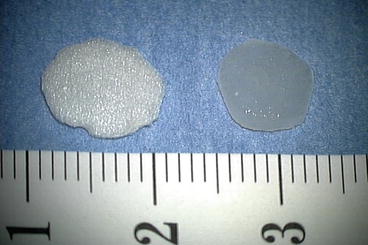
Fig. 4.1
Trimmed gelatin sponge and trimmed gelatin sponge with b-FGF
4.3 Therapeutic Procedures
The tympanic region was fully anesthetized by applying a cotton-ball soaked in 4 % lidocaine to the perforation in contact with the residual TM for 15 min. Mechanical disruption of the perforation edge was then created under the microscope by a myringotomy knife/micro-pick. A gelatin sponge that was larger than the perforation was immersed in b-FGF (5–30 μg of Trafermin (recombinant human basic FGF) of 100 μg/mL) then inserted into the perforation in contact with the perforation edge of the TM. Fibrin glue was then dripped over the sponge. Figure 4.2 shows these procedures in detail. In cases in which complete closure of the TMP was not achieved, the aforementioned treatment was repeated up to 4 times.
Fig. 4.2
Schematic diagram showing the method and procedures used in this treatment. 1 TM perforation. 2 Following local anesthesia with 4 % lidocaine, a mechanical disruption of the TM perforation edge is created under the microscope. 3 A gelatin sponge immersed in b-FGF is placed over the perforation in contact with the residual TM. 4 Fibrin glue is dripped over the sponge. 5 Three weeks after the treatment, residual crust is removed. In cases of incomplete closure of the TM perforation, the treatment is performed repeatedly
Patients were advised not to blow their nose and/or sniff strongly for a while and were ordered to revisit the hospital 3 weeks after treatment at which point any crust over the TM was removed.
The effectiveness of this treatment for TM repair was evaluated using certain indicators including closure rates, hearing level, sequelae, improvement of tinnitus, and aural fullness 3 weeks after treatment. The final evaluation was performed 3 months after finishing the treatment. Hearing levels were measured prior to and 3 months following the treatment. The success rate was determined based on the rate of complete closure of the TMP within four courses of treatment. In statistical analysis, Mann-Whiteny U test was used.
4.4 Results
4.4.1 Rates of Complete Closure of the TMPs
The overall rates of complete closure of the TMPs were 98.1 % (52/53) in the b-FGF group and 10.0 % (1/10) in the control group (Table 4.2). In the b-FGF group, the number/rate of the cases in which the TMP was able to close completely after one treatment, two treatments, three treatments, or four treatments were 41 (77.4 %), 7 (13.2 %), 3 (5.7 %), and 1 (1.9 %), respectively. There was only one case in which the TMP failed to close completely by the end of the fourth treatment. Figures 4.3 and 4.4 depict typical cases of perfect TM regeneration. In the control group, one patient with old traumatic TMP, classified as Grade I, showed complete closure of the TMP after three courses of treatment.
Table 4.2
Results
Grade | Total n = 53 | Grade I n = 9 | Grade II n = 25 | Grade III n = 19 |
|---|---|---|---|---|
Closure rates | 98.1 % (52/53) | 100 % (9/9) | 100 % (25/25) | 94.7 % (18/19) |
Number of treatment times | 1–4 (Avg.:1.4) | 1–3 (Avg.:1.3) | 1–4 (Avg.:1.4) | 1–4 (Avg.:1.6) |
Average HL Improvement | ||||
NA | 21.7 dB | 18.3 dB | 21.2 dB | 25.8 dB |
LA | 31.7 dB | 28.5 dB | 31.7 dB | 35.0 dB |
Adverse events | ||||
TO | n = 8 | n = 1 | n = 3 | n = 4 |
RTM | n = 6 | n = 0 | n = 3 | n = 3 |
Fig. 4.3
Case 1: A 89-year-old male with chronic otitis media persisting for 50 years. 1 Large, dry, Grade III perforation. 2 Disruption of the perforation edge. 3 Gelatin sponge with b-FGF was placed over the TMP and sealed by fibrin glue. 4 Three months after the treatment the TM was perfectly regenerated. Even in this elderly case, TM can be regenerated by this treatment
Fig. 4.4
Case 2: A 52-year-old male with otitis media with the TMP. 1 Large Grade II perforation: Almost all remaining TM was accompanied by calcification. 2 Two months after treatment, an almost normal TM with lots of blood capillary (white arrow) was regenerated. Remaining gelatin sponge (white triangle) was observed in the tympanic side. This will disappear within 3 or 4 months
4.4.2 Size of the TMP and Number of Treatments Required
Table 4.2 shows the number of treatments and the presence of any adverse events that occurred during the process of complete TMP repair for each grade of the b-FGF group.
4.4.3 Hearing Improvement
Pure tone audiometry was measured in all patients before and 3 months after treatment. Average hearing levels (average of 0.5, 1, 2 kHz) showed improvement in all patients with successful TM repair in the b-FGF group (Table 4.2). Hearing levels showed remarkable gains in the lower frequency ranges: 0.125, 0.25, and 0.5 kHz (Fig. 4.5).
Fig. 4.5
The change in hearing levels in the patient from case 1 (Fig. 4.3), 3 months after treatment with b-FGF. Hearing was measured before and 3 months after treatment. In this case, hearing recovery was mainly observed in the low-frequency ranges
4.4.4 Tinnitus and Aural Fullness
Prior to treatment, many patients had subjective symptoms. In the b-FGF group, 96.2 % (51/53) of the patients had tinnitus and 86.8 % (46/53) had aural fullness. Of these patients, 98.0 % (50/51) and 95.6 % (44/46) noticed improvements in tinnitus and aural fullness, respectively. In contrast, in the control group, 100 % (10/10) of the patients had tinnitus and 90% (9/10) had aural fullness. Of these, only 10 % (1/10) and 11.1 % (1/9) of the patients noticed improvements in tinnitus and aural fullness, respectively. Figure 4.6


Stay updated, free articles. Join our Telegram channel
Full access? Get Clinical Tree
