Ptosis surgery

CHAPTER 48 Ptosis surgery


Introduction


The term ‘ptosis’ is derived from the Greek word image, which means ‘to fall’. In ophthalmology and oculoplastics this term invariably and unequivocally refers to the pathologic condition in which the upper eyelid margin is inferiorly displaced. In the setting of a patient with ptosis, the abnormal eyelid position may cover a significant portion of the cornea and hence the pupillary aperture, sometimes to a degree that is enough to cause visual impairment from the obstruction created. A more suitable term to describe the condition would be that of blepharoptosis, but for the purposes of this chapter the terms ptosis and blepharoptosis will be used interchangeably.


The treatment of ptosis requires accurate patient evaluation, consistent measurement of the eyelid position and function, and careful documentation of the functional deficit. Skillful use of the various surgical techniques to implement a functional and esthetic correction is required from the performing surgeon.


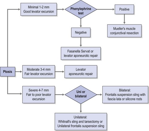
A practical clinical algorithm that allows the surgeon to select a reliable procedure for the management and treatment of the ptosis patient is provided. The algorithm’s goal is to help the surgeon with the selection of the best surgical procedure based on the amount of ptosis and levator function present the time of diagnosis, to achieve successful correction of the ptosis present with good postoperative results and patient satisfaction (Fig. 48.1).


Epidemiologic consideration and terminology


There is a uniform paucity in the literature of studies reporting the incidence and frequency of ptosis. There is no racial or gender predilection for either involutional or congenital ptosis occurring equally among the different races and between males and females.


The most frequent cause of ptosis is the dehiscence of the levator aponeurosis; therefore the incidence of involutional ptosis increases steadily with age. Blepharoptosis has been observed more frequently in soft contact lens wearers, and increasing years of contact lens wear seem to be associated with a higher rate of blepharoptosis of involutional nature. The frequency of involutional upper eyelid ptosis is difficult to determine, mainly due to its reversible morbidity and absolute lack of mortality. However, it is increasingly recognized in the elderly population. This is particularly true in patients who have undergone cataract extraction with or without lens replacement; the etiologic factor implied is perhaps the stretching or disruption of the levator palpebrae superioris muscle when the eye is maintained open for cataract surgery using a lid speculum.


Congenital ptosis is the second most frequent cause of blepharoptosis, while other causes of ptosis are relatively infrequent. A patient who develops an acquired ptosis over a period of days or weeks can signal a serious medical problem and needs further neurologic and physical evaluation.


Congenital ptosis can affect one or both eyes; however, in approximately 70% of known cases, congenital ptosis has unilateral affectation. Congenital ptosis may be present at birth, or it may develop later in life. A droopy eyelid(s) that is present at birth, or that develops within the first year of life, is considered congenital in nature. In most cases of congenital ptosis, the problem is isolated and does not affect the vision if the eyelid doesn’t obstruct the visual axis. This holds particularly true in the critical period of visual development, corresponding to the first 3 months of life. The pupil obstruction caused by the droopy eyelid obscures the pediatric patient’s visual field, and permanent loss of vision may occur as a result of amblyopia. Congenital ptosis has no mortality and its morbidity arises from occlusion (deprivation) amblyopia, astigmatism induced from the compression of the droopy eyelid on the corneal tissue, and ocular torticollis. The astigmatism induced by congenital ptosis is generally anisometropic and can be uni or bilateral depending on whether one or both eyelids are affected by the condition. In instances where the degree of anisometropia is significant, anisometropic amblyopia can also develop. In the presence of amblyopia and/or functional limitation caused by congenital ptosis, surgery must be performed to correct the problem early in life, since recent evidence supports that the incidence of deprivation or anisometropic amblyopia in congenital ptosis can be reduced significantly1.


Congenital ptosis can also be seen as part of many congenital syndromes including blepharophimosis syndrome, and certain forms of congenital fibrosis of the extraocular muscles syndromes (CFEOM)2, Marcus Gunn jaw winking syndrome, and congenital Horner syndrome


Clinical features, diagnosis, and differential diagnosis


Since a droopy upper eyelid is an obviously visible phenomenon, patients often present for consultation mainly due to cosmetic concerns. In fewer instances patients may also be aware of, and present because of, a decrease in superior visual field limiting their activities of daily life and often express an inability to read because of severe ptosis in downgaze. Based on its etiology, blepharoptosis can be classified as true ptosis or pseudoptosis. Pseudoptosis refers to the appearance of ptosis without true eyelid margin ptosis or levator dysfunction, and can be the cause of or associated with severe dermatochalasis, orbito palpebral asymmetry, and/or changes in both ocular and orbit volume. True ptosis could be congenital or acquired, and unilateral or bilateral. Ptosis describes an abnormally low and downward displacement upper eyelid in the relaxed primary position in relationship to its physiologic position 1 or 2 mm below the superior corneoscleral limbus. Regardless of whether the ptosis is congenital or acquired it can be classified based on its pathophysiologic mechanism as:


The upper eyelids are elevated by the contraction of the levator palpebrae superioris; with time the connections between the aponeurosis of the muscle and the tarsal plate, usually in older patients, suffer thinning, lengthening, or sometimes disinsertion of the levator aponeurosis from the tarsal plate, causing the eyelid to droop by these involutional changes. Acquired ptosis secondary to levator tendon disinsertion or aponeurotic dehiscence develops spontaneously, after episodes of allergic reactions, or after minor trauma including surgical trauma in older adults, as observed in some cases post-cataract extraction. The degree of ptosis that may develop after levator aponeurosis dehiscence might be minimal to moderate, ranging from only 1–1.5 mm of ptosis, to severe, with in some instances complete ptosis in which the patient is unable to open the eye. Ptosis of the eyelids can have a subtle presentation and even go unnoticed by the patient. In some cases, patients complain of restricted visual fields. In the ptotic eyelid, the lid crease is generally present and is often slightly higher than that of the uninvolved side. Clinically the levator excursion in this kind of ptosis is relatively good, and the ptosis remains essentially unchanged in both upgaze and downgaze; this differentiates it from the inelastic levator muscle typical of congenital ptosis. Presenting signs also include: persistent wrinkles in the forehead due to contraction of the frontalis muscle (in an unconscious attempt to elevate the eyelid), and asymmetric elevation of the eyebrows, greater on the affected side. Additionally, the upper sulcus is frequently deeper than on the uninvolved side, completing the picture for clinical diagnosis.


Acquired muscular dystrophy, progressive external ophthalmoplegia, and myasthenia gravis all can be causes of non-congenital, usually late onset, ptosis of myogenic origin. In these cases the pathology lies at the level of the neuromuscular junction producing an ineffective muscular contraction that is insufficient to keep the eyelids open and elevated. Myogenic ptosis usually points to a systemic etiology that requires medical management like myasthenia gravis, and should be fully worked up, using surgery only as the option to rehabilitate the patient, since treatment is almost palliative. Ptosis in the myogenic group is generally progressive and has a high frequency of recurrence despite repeated surgery, including levator resections. When a frontalis sling procedure is performed on these cases, surgery is usually limited by the development of exposure keratopathy and lagophthalmos. Patients with an adequate tear secretion and orbicularis muscle closure are considered to be the best candidates. Whenever possible, surgery should be performed in these patients under local anesthesia to allow intraoperative adjustment of the lid position, taking into consideration the degree of postoperative lagophthalmos that the individual can tolerate. Consideration of a surgical result aiming for under-correction in these patients can benefit certain cases.


The pathologic mechanism of acquired neurogenic ptosis differs from that of myogenic ptosis based on the fact that in neurogenic ptosis there is direct damage to the nerve(s) supplying the tone and innervation for the muscular contraction to occur, without affecting the motor endplate. These examples include acquired CN III palsy and damage to the cervical sympathetic nerve chain causing Horner syndrome.


Mechanical ptosis can occur due to a solid tumor, cyst, enlarged lacrimal gland, or any other ocular adnexal neoplasm pushing down the eyelid. The surgeon’s experience and knowledge regarding these conditions will dictate surgery that will address the causative problem, thus correcting the ptosis whenever possible.


Although traumatic ptosis legitimately points to an acquired form of ptosis, this kind of ptosis varies according to the location of the direct injury to the levator muscle or its neurovascular supply. Mild degrees of trauma, whether or not associated with edema or hemorrhage, can lead to levator aponeurosis disinsertion that can be repaired using an aponeurotic approach surgery. Lacerations of the lid may sever the soft tissues, leading to scarring and secondary mechanical ptosis. When possible this problem is best managed by early and meticulous repair of the disrupted levator aponeurosis at the time of primary repair of the lid injury. When primary repair is not possible, often the eyelid and the orbit can be explored at a later time and the levator muscle identified and repaired, although the technical challenges associated with a delayed repair sometimes render suboptimal results even in the best of experienced hands. In certain instances traumatic ptosis involves damage to the nerve supply of the levator muscle. Since the levator and the superior rectus muscle share a common innervation, these injuries may affect the eye elevation, sometimes even limiting the globe’s upward excursion during Bell’s phenomenon. It is not uncommon for the patient to experience some degree of neural regeneration after trauma, so it is not a bad idea to wait at least 6 months to 1 year prior to any surgery to allow some degree of spontaneous regeneration to happen. After this period, when the degree of recovery has reached its maximum point, it is reasonable to attempt a levator resection; or a sling procedure can be performed, depending on the severity of ptosis and the degree of return of levator muscle excursion.


Although there are numerous classifications for ptosis; such as congenital versus acquired, neurogenic, myogenic, traumatic, and mechanical3, none of those classifications provides a complete approach to the ptosis patient, nor does any of them guide the clinician to the development of a system for adequate repair. Classifying ptosis as minimal = 0.5–1.5 mm, moderate = 2.0–3 mm, or severe >3 mm based on the amount of ptosis provides a practical framework for a logical system of repair when this system is used in conjunction with the amount of levator excursion. This last measurement is an indirect measurement of the viability of the levator palpebrae superioris and an excellent predictor of the surgical outcome. Using this matrix the appropriate choice of surgical strategy can then be applied to each of these three scenarios.


Goals of surgery


Blepharoptosis is one of the most commonly encountered oculoplastic problems, but also at the same time one of the most challenging problems from the surgical standpoint. The goal of ptosis surgery is to obtain, as much as possible, an anatomic and physiologic result by elevating the lid or lids to an adequate position and, if necessary, to clear the pupil from the obstruction created by the eyelid’s downward displacement. Additionally, special attention must be given not to disrupt the natural contour and symmetry of the eyelids.


Thorough understanding of these goals and of the limitations of ptosis surgery is important, for both the surgeon and the patient. Also patients and their families must be fully aware of these goals and limitation before surgery is performed. It is advisable that the expectations and anticipated surgical results are discussed carefully with the patient and/or the parents preoperatively and documented in writing as part of the informed consent process. This proves particularly true in congenital ptosis, since in this instance factors inherent to the anatomic defect of the defective levator palpebrae superioris muscle impose limitations on the surgical results. Surgery cannot restore the function to a compromised muscle with an already abnormal or absent function preoperatively. In congenital ptosis with a frontalis suspension procedure or a maximal levator resection the lid level can be changed, but dynamic results will not be obtained postoperatively, and surgery may result in significant lid lag and lagophthalmos that is still more obvious on downgaze. The best result that can be hoped for in congenital ptosis surgery is a normal lid level, contour, and symmetry when the eyes are in the primary position, with adequate clearance of the visual axis.


Indications for surgery


In patients with acquired blepharoptosis, surgery is often recommended when the patient’s activities of daily life are compromised by the occlusion of the visual axis caused by the droopy eyelid, a significant superior visual field is reported or perceived by the patient, or extreme fatigability of the eyelid and occlusion of the pupil occur while in downgaze, affecting mainly activities like reading amongst others. Vision may be affected in patients regardless of the amount of ptosis. During the preoperative evaluation, visual or asthenopic symptoms secondary to the ptosis might be elicited; when these are present, they usually indicate the need for surgery. Third-party payers now require documentation of symptoms, field defects, and by clinical photographs routinely.


In most instances, the primary reason prompting the parents to seek for help in correcting congenital ptosis is cosmetic. It is generally agreed that disfiguring congenital ptosis should be repaired by age 5 years or before the child begins regular school to avoid psychosocial issues during the child’s socialization phase, but setting an arbitrary age for surgery serves no purpose. Earlier intervention is performed in children with severe bilateral ptosis that interferes with the child’s ability to learn how to walk, due to the extreme head position that they adopt with the chin-up position. Another exception arguing for early intervention is made in patients with unilateral or bilateral severe congenital ptosis where the normal visual development is compromised by total occlusion of the visual axis. In some of these instances surgical intervention may be even indicated shortly after birth. Disruption or loss of compensatory mechanisms of binocular fusion such as chin-up head position is a sign of development of amblyopia, which must be treated urgently with surgical correction of the ptosis and appropriate amblyopia management, with occlusion therapy and glasses when necessary.


Preoperative assessment


In the evaluation of the ptosis patient, history taking is one of the most important elements. If the ptosis is congenital, the physician should question the patient or family about the absence or presence of jaw winking and family history of ptosis. With acquired ptosis, the timing of onset is of extreme importance (acute vs. progressive or chronic). A history of fatigability or variable ptosis with exercise or through the day should warrant a work-up for myasthenia gravis, especially if there is noticeable improvement with rest. Associated neurologic symptoms, if any, should also be investigated. History of orbital or ocular trauma, previous ocular histories of inflammatory disorders, or contact lens wear may also be germane. The ocular exam should be complete. In addition to documenting the patient’s visual acuity and ocular motility, the importance of the pupillary function exam should not be underestimated. Anisocoria suggestive of Horner syndrome should be fully evaluated. The presence or absence of Bell’s phenomenon should be documented, as well as quantitative and qualitative properties of the tear film both by Schirmer’s testing and by study of tear break-up time. Slit-lamp examination should be used to assess the integrity of the cornea and the conjunctiva and to detect any inflammatory disease or subclinical pathology.


Children with ptosis should have full-dilated exams, retinoscopy, and assessment of ocular motility and sensory function with either fixation preference, the 10Δ base-down prism test, or a formal stereopsis test to rule out amblyopia. Careful motility examination in pediatric patients may uncover a double elevator palsy associated with the ptosis or partial third cranial nerve involvement causing the ptosis.


Assessment of ptosis and documentation


Ptosis is documented by the margin to reflex distance 1 (MRD1)4, which is the distance from the central pupillary light reflex to the upper eyelid margin, measured in millimeters. It is important to document the amount of ptosis to the nearest 0.5 mm, if possible. The margin to reflex distance 2 (MRD2) is the distance from the central pupillary light reflex to the lower eyelid margin. The MRD1 plus the MRD2 should equal the palpebral fissure.


The levator excursion test is the best clinical means for assessing levator function. The levator excursion is documented in millimeters, measuring the distance from extreme upgaze to downgaze with the brow immobilized by the examiner’s thumb to eliminate any contribution of the brow to lid elevation. A millimeter ruler is used vertically in the pupillary axis to assess the full excursion. Levator excursion of 10 mm or greater is considered good function; 5–9 mm of excursion is fair function; and 4 mm or less is poor function.


Patients with minimal ptosis (2 mm or less) should have a phenylephrine test performed in the involved eye or eyes after appropriate ptosis measurements have been documented. Either 2.5% or 10% phenylephrine is instilled in the affected eye or eyes. We prefer to use 2.5% phenylephrine. Usually two drops are instilled and the patient is re-examined 5 minutes later. The MRD1 is rechecked in the affected and unaffected eyes (Fig. 48.2). A rise in the MRD1 of 1.5 mm or greater is considered a positive phenylephrine test. This indicates that Müller’s muscle is viable, and the Müller’s muscle conjunctival resection procedure can be performed, also giving the patient a reasonable prediction of the desired result.


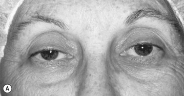
The contralateral eye must also be rechecked in patients with unilateral ptosis. When the ptotic eye is occluded, if the MRD1 decreases appreciably in the opposite eye, this usually indicates that bilateral ptosis is present; this finding is consistent with Hering’s law5. The patient may require bilateral surgery. A negative phenylephrine test precludes the use of the Müller’s muscle conjunctival resection procedure, because the outcome of the procedure is unpredictable in this setting. Callahan and Beard3

Only gold members can continue reading. Log In or Register to continue

Stay updated, free articles. Join our Telegram channel

Jun 4, 2016 | Posted by in OPHTHALMOLOGY | Comments Off on Ptosis surgery

Full access? Get Clinical Tree

Get Clinical Tree app for offline access