Figure 13.1  Silicone sponges bulge between mattress sutures at the end of buckle, which causes conjunctival erosion (dellen formation), which leads to exposure of buckle and extrusion. In addition, buckle is flexible, which results in a lower buckle between mattress sutures, leading to radial folds and unsupported retinal breaks.
Implants Versus Explants
While scleral dissection is an acceptable method of scleral buckling, it is rarely performed currently because of its inflexible, time-consuming nature. The original justification for scleral dissection was the ability to avoid scleral damage from full-thickness diathermy. Although burying the buckle under a flp reduces extrusion, it increases intrusion, operating time, inflexibility, and the risk of scleral perforation.
Buckling with Vitreous Surgery
Scleral buckling is used with many variations for retinal reattachment (13,14). A rapid, simplified form of scleral buckling is preferred by the authors for all scleral buckling (Fig. 13.2). Scleral buckling is significantly overutilized in conjunction with vitreous surgery. Scleral buckling causes a very significant incidence of strabismus, ptosis, pain, conjunctival damage, and refractive error. Scleral buckling is not indicated for giant break surgery, PVR, or routine rhegmatogenous retinal detachments. The elimination of vitreous traction coupled with complete intraoperative reattachment by vitrectomy has eliminated, in the authors’ opinion, the need for combined buckle-vitrectomy.
Figure 13.2  A simplified approach saves time, reduces complexity, and reduces complications.
Encircling Bands
Prophylactic scleral buckling with an encircling band can then be thought of as making a new ora serrata to treat peripheral vitreoretinal traction preceding retinal detachment. Encircling bands for prophylactic buckling were used frequently in the early days of vitrectomy (15). This approach is not used today because of better cutters, techniques, fluidics, and dissection methods. Local anesthesia, outpatient surgery, cost containment as well as avoidable complications such as pain, strabismus, refractive error, conjunctival damage, and ptosis all contribute to the virtual elimination of prophylactic buckling.
Circumferential Explants
Because of their narrow configuration, bands alone are usually not utilized to treat specific retinal breaks. If a circumferential explant can cover the posterior extent of a retinal break, it is utilized in preference to a radial explant. Circumferential explants require less exacting localization, do not distort the macula, and cover a broader extent of vitreous base pathology. Posterior breaks are managed with vitrectomy techniques. The principal author has not used radial buckles or sponges for over 25 years in the buckling alone or vitrectomy setting.
Monofilament (5-0) nylon sutures are utilized with a single circumferential posterior scleral bite. In contrast to radial suture bites, the circumferential bite can be quite long without reducing the posterior extent of the buckle. The single circumferential posterior scleral bite reduces by one half the chances of perforating the retina, as compared to paired bites. This posterior bite is always placed 3 mm and preferably 5 mm posterior to the most posterior aspect of the most posterior break. A too-anterior positioning of the buckle causes many reoperations after scleral buckling procedures.
All anterior scleral bites are placed circumferentially in the scleral condensation, conforming to the rectus muscle insertions. This provides an area of thicker sclera for greater permanence. This muscle ring also conforms to the ora serrata; therefore, a scleral suture bite placed here in a circumferential orientation cannot perforate the retina (Fig. 13.3). Extending all circumferential buckles to the ora serrata prevents the anterior leakage of subretinal fluid (SRF) associated with narrow bands or buckles placed more posteriorly.
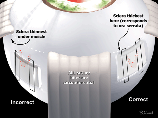
Figure 13.3  Anterior bites of mattress suture should be made in thicker sclera associated with the rectus muscle insertions. The thicker sclera reduces postoperative pull-through, and the location corresponds to the ora so that perforation will not damage the retina. Placing all sutures this anterior prevents SRF from leaking anteriorly.
Stay updated, free articles. Join our Telegram channel

Full access? Get Clinical Tree


