The Three Germ Layers: Ectoderm, Endoderm, Mesoderm
The auricle is innervated by three nerves:
• The great auricular nerve (originating from the cervical plexus). It supplies the descending helix and the lobule. Here are the reflex zones of organs derived from the ectoderm (skin, nervous system).
• The auriculotemporal nerve (a branch of the trigeminal nerve). It supplies the ascending helix and superior helix as far as Darwin’s tubercle, the triangular fossa, the scapha, the antitragus, and the antihelix including the antihelical wall. Organs derived from the mesoderm are projected in this region of the ear (muscles, bones, ligaments, heart, kidney).
• The auricular branch of the vagus nerve. It supplies the concha—the projection zone of organs derived from the endoderm (internal organs, with the exception of kidney and heart). This zone extends to the projection zone of the sympathetic trunk where the concha merges into the antihelical wall.
This pattern of auricular innervation is easy to memorize and will be of great help when learning the reflex zones of the ear. Each of the three areas of innervation corresponds to one of the three germ layers (ectoderm, mesoderm, and endoderm) and is thus assigned to a clearly defined group of organs.
Remarkably, the representation of organs on the auricle follows this pattern in almost all cases—for example, the Kidney Point does not lie in the zone of the endoderm like the reflex points of other internal organs; it is found beneath the ascending helix in the zone of the mesoderm from which the kidney is derived.
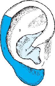
Fig. 2.1 The three germ layers
1 Ectoderm
2 Endoderm
3 Mesoderm
Locomotor System—Overview
The locomotor system projects onto the upper terrace of the ear (scapha, triangular fossa). In principle, the sensory points lie on the front of the ear (lateral surface), while the motor points are on the back of the ear (medial surface). The forceps method according to Bahr is based on this arrangement:
A gold needle in the sensory point on the lateral surface, and a silver needle in the corresponding motor point on the medial surface.
Body parts, organs, or anatomical structures belonging to the same region project very close to each other onto the ear. For example: knee joint, popliteal artery, peroneal nerve.
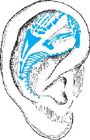
Fig. 2.2 Projection of the locomotor system
Entire Vertebral Column
Location:
• The vertebral column, or spine, projects upside-down onto the entire antihelix.
• Start: Postantitragal fossa (Atlanto-occipital Joint Point, Point C0/C1).
• End: Coccyx Point at the end of the antihelix, underneath the ascending helix (only visible with the helix unfolded; very close to the Internal Anus Point). The outer aspect of the antihelical fold, or reflection (scapha-antihelical wall), is variably expressed, which helps in subdividing the zone of the vertebral column into Cervical Spine Zone, Thoracic Spine Zone, and Lumbar Spine Zone.
Structures of the Vertebral Column
Location:
• Like the various structures of a vertebra, the corresponding points lie close together on the edge of the antihelix:
• The small vertebral joints project slightly lateral to the edge of the antihelix (which is important for treating the very common “blockages” of the spine). The Zone of Paravertebral Muscles and Ligaments lies lateral to this Zone of Small Vertebral Joints.
• Points nearby: At the level of the transition from the upper third to the lower two thirds of the antihelical wall lies the Zone of Nervous Control Points of Endocrine Glands, at the foot of the antihelical wall lies the Zone of Sympathetic Trunk (Zone of Paravertebral Chain of Sympathetic Ganglia) with the reflex points for the nervous control of organs (e.g., Nervous Liver Point) and for the cervical ganglia (e.g., Stellate Ganglion Point). See Nervous System, page 173.
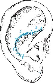
Fig. 2.3
1 Cervical spine
2 Thoracic spine
3 Lumbar spine
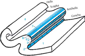
Fig. 2.4
1 Zone of Parenchymal Organs (Concha)
2 Zone of Sympathetic Trunk, Neural Organ Points
3 Zone of Nervous Control Points of Endocrine Glands
4 Zone of Intervertebral Disks
5 Zone of Vertebrae
6 Zone of Small Vertebral Joints
7 Zone of Paravertebral Muscles and Ligaments
8 Zone of Spinal Cord
Cervical Spine
Location:
• In the inferior portion of the antihelix (Zone of Cervical Spine), the antihelical fold is sharply defined toward the conchal wall, like a tube with a small diameter.
• At the level of Point C7/T1, there is a distinct change: the sharp edge widens and becomes gently rounded (Zone of Thoracic Spine). The curvature of the antihelix also changes.
Thoracic Spine
Location:
• The zone between Points C7 and L1, with the antihelix being gently rounded (a sharp edge in the Cervical Spine Zone, a steep cartilage overhang in the Lumbar Spine Zone; see above and next page).
• Relative to the total length of the thoracic spine in the body, the segment of its representation on the ear is short.
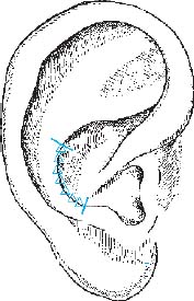
Fig. 2.5 Cervical spine
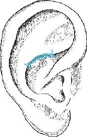
Fig. 2.6 Thoracic spine
Lumbar Spine
Location:
• Start: About 1cm before the antihelix intersects the ascending helix. The soft rounding of the Thoracic Spine Zone abruptly changes into a steep or lamella-shaped cartilage overhang. The latter represents the Lumbar Spine Zone.
• End: Point L5/S1 at the intersection of antihelix and helix. From here on, underneath the ascending helix, the sacral vertebrae are projected.
C0/C1 (Atlanto-occipital Joint)
Location:
• Postantitragal fossa, easily detectable with the stirrup-shaped ear probe (see also Cervical Spine, p. 15).
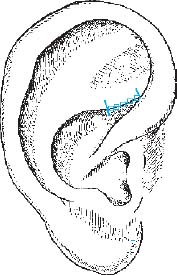
Fig. 2.7 Lumbar spine
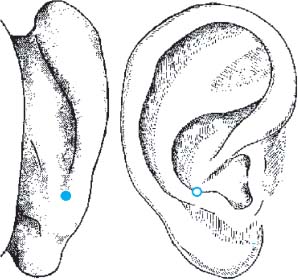
Fig. 2.8 C0/C1 (atlanto-occipital joint)
C7/T1
Location:
• Where the sharp edge of the Cervical Spine Zone turns into a gentle rounding. Easy to find with the stirrup-shaped ear probe (see also Cervical Spine and Thoracic Spine, p. 15).
T12/L1
Location:
• Where the gentle rounding of the Thoracic Spine Zone turns into a sharp, overhanging edge. Easy to find with the stirrup-shaped ear probe (see also Thoracic Spine and Lumbar Spine, pp. 15 and 17).
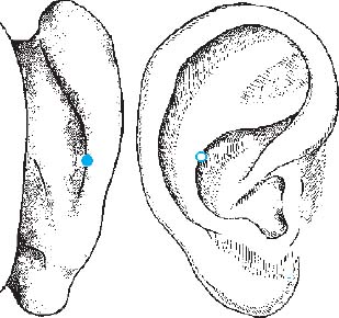
Fig. 2.9 C7/T1
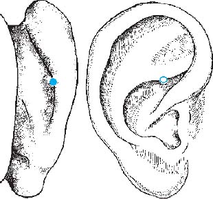
Fig. 2.10 T12/L1
L5/S1 (Lumbosacral Transition)
Location:
• On the antihelix, at the intersection with the ascending helix (see also Lumbar Spine, p. 17).
Coccyx
Location:
• At the end point of the antihelix, underneath the ascending helix. Easily palpable with the finger. All points of the sacrum lie on the antihelix hidden beneath the ascending helix.
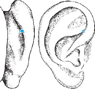
Fig. 2.11 L5/S1 (lumbosacral transition)

Fig. 2.12 Coccyx
Sacral Spine
Location:
• The sacrum projects onto the part of the antihelix that runs beneath the ascending helix.
• Start:
Stay updated, free articles. Join our Telegram channel

Full access? Get Clinical Tree


