Head—Overview
The reflex areas of the head are localized on the lobule and the descending helix. They include the projections of the nose, eyes, ears, tongue, teeth, tonsils, lymph nodes, maxillary sinuses, and frontal sinuses.
Also the brain projects onto the lobule; it is discussed under Nervous System (p. 121).
The entire head projection deviates from all other reflex areas on the ear. Instead of the usual upside-down arrangement, the head organs project in about the same arrangement as in an upright-standing person.
The upright arrangement of the head’s reflex zones becomes clear when imagining the famous “inverted embryo” in an orientation similar to the face presentation within the uterus. In this position, the lower jaw necessarily projects caudally, not cranially, relative to the upper jaw. Likewise, the nose projects caudally to the mucous portion of the paranasal sinuses.
Bony Skull
Location:
• On the outer surface of the antitragus. The reflex area of the occipital bone (Occiput Point) borders immediately on that of the uppermost cervical vertebra (Point C1 or Atlas Point).
The postantitragal fossa forms the boundary between the projections of the uppermost cervical vertebra (Point C1) and the base of the skull. Here is the reflex area of the atlanto-occipital joint (Point C0/C1).
The other cranial bones project anteriorly and inferiorly to the occipital bone:
• the frontal bone and sphenoid bone in the anterior third,
• the parietal bone and temporal bone in the middle third, and
• the occipital bone in the posterior third of the zone.
The reflex area of the maxilla (Upper Jaw Zone) forms the caudal border of the reflex zone of the cranial bones.
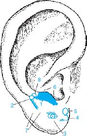
Fig. 4.1
1 Upper and lower jaws
2 Projection of throat, palatine tonsil, pharyngeal tonsil, and lymphoid ring
3 Eye
4 Nose
5 Maxillary and frontal sinuses
6 Bony skull
7 Tongue
8 Ear
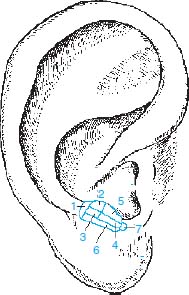
Fig. 4.2
1 Occipital bone
2 Parietal bone
3 Temporal bone
4 Sphenoid bone
5 Frontal bone (Forehead Point)
6 Maxilla
7 Frontal sinus
Temporomandibular Joint
Location:
• At the end of the scapha where the helical groove merges into the lobule.
The following points are located very close to the Temporomandibular Joint (TMJ) Point and therefore indistinguishable from it when starting the diagnosis:
• Palatine Tonsil Point,
• Molar Point (for the molars of upper and lower jaws),
• Retromolar Cavity Point,
• Posterior Masseter Muscle Point (sensory portion),
• Depression Point, and
• Magnesium Point.
At the same location are also the points for
• the central portion of the deep and superficial parotid gland (affected only occasionally), and
• the insertion of the lateral pterygoid muscle (which adducts discus and lower jaw, causes tenseness during bruxism).
To distinguish between these reflex areas, distinct points of body acupuncture may be enlisted, or the assignment on the head or face may be made by means of the VAS.
See also B. Strittmatter, Overcoming Blockages to Healing, Thieme Publishers, Stuttgart–New York, 2004.
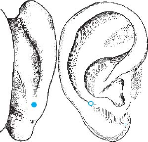
Fig. 4.3 Temporomandibular joint
Upper and Lower Jaws, Teeth
Location:
•
Stay updated, free articles. Join our Telegram channel

Full access? Get Clinical Tree


