Key points
- •
Facial nerve schwannomas are the most common primary tumor of the facial nerve and represent a difficult treatment paradigm.
- •
There is significant debate over optimal treatment of facial nerve schwannomas with observation, decompression, debulking, resection with facial nerve grafting, and radiosurgery all supported in the literature.
- •
Geniculate ganglion hemangiomas often present with facial nerve paralysis out of proportion to lesion size on imaging.
- •
Treatment of geniculate ganglion hemangiomas has changed over time with earlier surgical resection playing a more modern role.
| CPA | Cerebellopontine angle |
| FN | Facial nerve |
| FNS | Facial nerve schwannoma |
| GG | Geniculate ganglion |
| GGH | Geniculate ganglion hemangioma |
| HB | House-Brackmann |
| IAC | Internal auditory canal |
| SRS | Stereotactic radiosurgery |
| VS | Vestibular schwannoma |
Introduction
Primary tumors of the facial nerve (FN) are rare with FN schwannomas (FNSs) being the most common of these lesions. Other less frequent tumors include glomus facialis, geniculate ganglion hemangiomas (GGHs), and granular cell tumors. Diagnosis of these lesions can be difficult given the intimate anatomic relationship of these tumors with other structures of the lateral skull base. Treatment is also challenging because the risk of FN injury is high with surgical intervention. Also, given the scarcity of these lesions, it is difficult to develop a clear consensus with regard to treatment. This article discusses the pathophysiology, symptomology, diagnosis, and treatment of these tumors.
Facial nerve schwannomas
FNS represents one of the more difficult treatment paradigms in neurotology. These tumors are so rare that determining a true incidence in the general population is difficult. Saito and Baxter found 5 (0.83%) incidental FNS in 600 temporal bone specimens representing the best current estimation. However, this collection is not representative of the general population and the actual incidence of FNS remains relatively unknown. This low case prevalence has made development of a uniform treatment algorithm challenging.
Presentation
The presenting symptoms of FNS vary based on tumor size and location. In patients ultimately diagnosed with FNS, hearing loss has been reported in 33% to 78.6%, tinnitus in 7% to 51.8%, and vertigo in 46%. Specifically with regard to FN function, 50% to 60% have some history of FN paralysis with approximately 20% of symptoms being transient.
Diagnosis
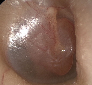
The patient’s physical examination is typically normal with a few exceptions. A thorough FN examination is important to identify any paralysis, twitching, or subtle abnormalities. The otologic examination is typically normal unless the tympanic or mastoid segment is largely involved revealing either a retrotympanic or external auditory canal mass ( Fig. 1 ). In such situations tuning fork examination can reveal a conductive loss on the involved side. Similarly, when there is a significant sensorineural hearing loss from an FNS of the internal auditory canal (IAC)/cerebellopontine angle (CPA), the Weber can lateralize to the uninvolved side.
The patient’s collective symptoms typically lead to imaging, which is the main diagnostic test for FNS. MRI and computed tomography (CT) can be helpful in diagnosing these lesions. Thin-sectioned MRI with gadolinium can positively identify an enhancing lesion along the FN. When segments proximal and distal to the Geniculate ganglion (GG) are involved, the typical “hourglass” appearance can be observed on the MRI ( Fig. 2 ). However, differentiating FNS from vestibular schwannomas (VS) can be difficult, if not impossible, when only segments proximal to the labyrinthine portion are involved. Likewise, if only the GG, tympanic, and/or vertical segments of the FN are involved, diagnosis can be difficult because these segments can also enhance under normal physiologic conditions. CT can complement MRI in looking at changes to the bony architecture surrounding the FN in the tympanic and mastoid segment, typically showing an enlarged fallopian canal compared with the contralateral side ( Fig. 3 ).
FNS can occur at any point along the FN. The most commonly involved segment is the GG (60%–66%) followed by the tympanic (53%) and labyrinthine segments (50.6%–60%). Most tumors involve more than one segment. Skip lesions can be seen on MRI when normal FN exists between involved segments. This has been described as “beads on a string” and has been reported to occur in 20% of cases.
Like their vestibular counterparts, FNS arise from the Schwann cells and are arranged in an Antoni A, Antoni B, or combined histologic pattern. There has been no described correlation between histologic pattern and tumor behavior.
Treatment
Determining the optimal treatment of an individual patient with an FNS is difficult. The overall goal of FNS treatment is preservation of FN function for the longest duration possible assuming other symptoms do not require intervention. Examination of the literature shows a wide variety of treatment methods. These range from conservative management with periodic imaging, decompression, tumor debulking or stripping, and resection with FN graft if possible.
One of the main difficulties in diagnosing and treating FNS is that many are initially thought to be VS until the tumor is exposed in surgery. This is particularly true when only the IAC and CPA are involved with tumor. Rates of FNS thought preoperatively to be VS range from 22% to 64.2%. Given this high rate, the first treatment decision has often already been made based on the presumption of being a VS. As MRI resolution continues to improve, it is hoped this rate will decline.
Like VS, FNS are slow-growing lesions. Rates of growth have been reported to be between 0.85 and 1.4 mm per year. As such, most authors recommend conservative management with serial imaging when symptoms are minimal and the FN function remains better than House-Brackmann (HB) grade III. In several reports, FNSs have been followed for more than 5 years before requiring intervention. Those patients who never required intervention have been able to maintain their baseline HB function with no impact on hearing. This is the ideal patient outcome, but unfortunately not always possible because of tumor growth and worsening symptoms.
Angeli and Brackmann introduced FNS decompression as a more conservative surgical treatment to preserve FN integrity and relieve pressure to allow improved axonal flow ( Fig. 4 ). This seems to be an effective measure to allow FNS to continue to grow, but at a slow pace and with less neuronal injury. Multiple patients have been able to be treated with decompression without significant decreases in FN function over time.
Wilkinson and colleagues reported 19 patients treated with FN decompression with a mean 6-year follow-up. Four patients (21.0%) showed a decline in FN function by one HB grade over time and three showed improved HB grade. There was also an increase in pure tone average by a mean 4.0 dB over this time period. These patients also showed decreased FNS growth rates of 0.17 mm per year compared with 0.85 mm per year in the observation cohort. Similar results from the University of Iowa showed continued normal FN function 2 years after FNS decompression. FN decompression has become a reasonable option in patients with growing bone-confined FNS and worsening FN function.
Although first introduced by Pulec in 1972, FNS tumor debulking remains controversial. The histologic evidence of FN fibers running throughout FNS in tumor specimens has caused some to reject this treatment method. Nevertheless there are numerous reports in the literature showing good FN outcomes using debulking for FNS. Supporters of debulking surgery believe that given advances in FN monitoring technology, portions of tumor that do not directly stimulate can be removed until fibrillation potentials are seen. Unfortunately, FN stimulation at the end of the case has not been shown to be predictive of FN outcome in these cases. Based on tumor size and location, patient symptomology, and preoperative hearing, FNS debulking can be performed either through a middle cranial fossa, transmastoid, translabyrinthine, or combined approach.
Mowry and colleagues reported on 11 patients with FNS isolated to IAC and CPA that underwent debulking. They were able to perform 95% tumor removal in nine patients (81.8%), 80% removal in one (9.1%), and 66% tumor removal in one (9.1%). One patient who was lost to follow-up had a preoperative HB grade I but developed an HB grade IV in the immediate postoperative period. Otherwise, multiple patients who preoperatively had HB grade III or worse improved their FN function to an HB grade I to II after surgery. Only one patient (10%) developed an HB grade III with a mean follow-up of 28.2 months (range, 2–96 months). One patient did develop tumor recurrence that required complete resection of the FNS with an FN graft.
In a recent study, Park and colleagues attempted to identify factors to determine the best candidates for debulking surgery. Of the 28 patients undergoing surgery for FNS, 18 (64.3%) underwent FNS debulking. Of these 18 patients, two (11.1%) developed worse than HB grade III function. They identified several factors that they believed allowed them to perform tumor debulking with minimal postoperative FN injury. Patients with good outcomes, considered HB grade I to II after debulking, were more likely to have tumors less than 2 cm; two or less involved FN segments, and had tumors proximal to and including the GG. Because of the small sample size, however, none of these factors reach statistical significance. One patient (5.6%) had continued growth of residual tumor and required stereotactic radiation therapy.
Data on subtotal tumor resection are sparse, but do show that a subset of patients can maintain good FN function postoperatively. Long-term follow-up is required to determine the viability of this option for FNS treatment. In the paper with the longest follow-up (mean, 7 years), Li and colleagues reported they were able to perform 95% tumor resection in 10 patients and 70% to 80% resection in five patients. Overall, 27% of patient showed tumor growth over time. This rate increased to 60% when looking only at those undergoing 70% to 80% debulking. Although the sample size was small there was a clear trend toward increased likelihood of growth in those undergoing smaller percentage resections. This is helpful in counseling patients after surgery, but does not help in the treatment decision-making process because it is impossible to know the extent of safe tumor debulking before surgery.
Patients presenting with poor FN function (>HB grade III) are less difficult treatment decisions. In this group with enlarging tumors, complete resection of the FNS with FN graft is the best option. For this population, outcome expectation should be lowered because the best possible outcome with an FN graft is HB grade III. In such cases, FN function has been shown to be improved or the same in 52% to 55% of patients, with 84% having HB grade III or IV. Collagen nerve tubules have been used recently and may help improve FN outcomes in this population. Difficulty arises in these cases, however, when the FN is found to be involved at the brainstem, without a proximal stump, and is thus not amenable to grafting. In such cases, FN reanimation procedures are valuable.
Independent studies by the authors have shown a substantial change in the management of FNS over time. Data from the House Ear Clinic show that before 1995, 85% of diagnosed FNS underwent resection, 15% decompression, and 0% observation. In contrast, in those diagnosed after 1995 only 27% underwent resection, 32.7% decompression, and 29% observation. A similar pattern of more conservative management was also reported from the Otology Group of Vanderbilt. Here the number of FNS resections with graft decreased from 74% to 40% and the number of subtotal resections increased from 2.1% to 30% over a similar time period. As the understanding of these tumors improves, the treatment algorithm is likely to continue to evolve.
A more recent addition to the management of FNS is the application of stereotactic radiosurgery (SRS). Unfortunately, like VS SRS literature, early reports on its effectiveness used vague terms for tumor control, hearing, balance, and FN outcomes. Recently, standard outcomes measures have been used with increased frequency. The full range of SRS techniques have been used to treat FNS with tumor control rates ranging from 83.3% to 100%. In most patients, FN function has remained unchanged, but there are multiple reports of FN function worsening after treatment.
One of the main factors that make understanding the outcomes of SRS for FNS difficult is that a large percentage of patients in the literature had surgery before their radiation therapy where the FNS was initially thought to be a VS. Presumptively, these patients then underwent decompression surgeries without tumor resection. We have previously discussed that FNSs undergoing decompression show slower rates of growth. Likewise, FN injury from post-SRS tumor swelling is presumably less likely if a decompression is performed. Therefore, separating the outcomes in those previously treated with surgery and not is difficult given the small patient population. Long-term data are needed to determine the efficacy of this treatment.
Stay updated, free articles. Join our Telegram channel

Full access? Get Clinical Tree


