Purpose
To estimate the age- and sex-specific prevalence of early age-related macular degeneration (AMD; drusen and retinal pigment abnormalities) and late AMD (exudative AMD and geographic atrophy) in the Japanese population.
Design
Community-based, cross-sectional study.
Methods
The study was held in Nagahama, Japan, and included 6065 Japanese individuals (aged ≥50 years) recruited in 2008-2010. We graded fundus photographs of both eyes for the AMD phenotype based on drusen size, the presence of retinal pigment abnormalities, and late AMD. The associations between smoking and AMD phenotypes were also evaluated.
Results
We assessed 5595 subjects (women, 65%) with a gradable macular condition. Early and late AMD prevalence increased from 16.1% and 0.27% at 50-59 years to 31.2% and 0.98%, respectively, at 70-74 years and was predominant in male subjects in each age group. Smoking was associated with both early and late AMD stages and retinal pigment abnormalities ( P < .0001), but not with drusen ( P = .305). The prevalence of retinal pigment abnormalities was significantly higher in men ( P < .0001), which was associated with high rates of cigarette smoking. We found no sex difference for the prevalence of large drusen ( P = .264).
Conclusions
The prevalence of early AMD among adult Japanese persons was similar to the rates in white populations. The prevalence of late AMD in Japanese people aged <70 years was similar to that observed in white populations, whereas that in Japanese people aged ≥70 years was relatively lower.
Age-related macular degeneration (AMD) is the leading cause of visual impairment in the elderly and is the most common cause of blindness in developed countries. The stages of AMD are categorized as early, in which visual symptoms are inconspicuous, and late, in which severe vision loss is typical. Early AMD is characterized by drusen or by pigment abnormalities of the retinal pigment epithelium (RPE) in the macula, without visible choroidal vessels. The presence or absence of these 2 features is characteristic of AMD and is highly associated with the development of late AMD, especially when the status of both eyes is considered.
To date, the introduction of anti–vascular endothelial growth factor (VEGF) intravitreal injections has offered remarkable clinical benefits for patients with late AMD. However, because these benefits are associated with an increased financial burden of providing care for these patients, determining the precise incidence of AMD and identifying its risk factors are still required in order to develop preventive measures for this disease. In fact, an increasing number of studies have reported the epidemiology of AMD in different racial/ethnic groups over the last 10 years. However, although the state of health, food intake, nutritional intake, and lifestyle of the Japanese people have been changing, only 2 small cohorts, the Hisayama study (1998), with 1486 participants aged ≥50 years, and the Funagata study (2000-2002), with 1246 participants aged ≥50 years, have evaluated the prevalence of AMD in the Japanese population.
These 2 population-based studies (the Hisayama study and Funagata study) arrived at similar conclusions regarding the prevalence of late AMD: late AMD is less common among Japanese people (with a reported overall prevalence of 0.87% and 0.6%, respectively) than among white subjects. However, these 2 studies arrived at different conclusions regarding the prevalence of early AMD in Japanese. Although the Hisayama study suggested a lower prevalence of early AMD in the Japanese, the Funagata study indicated that the prevalence of early AMD is similar to that reported in the Blue Mountains Eye Study (BMES). A recent meta-analysis in 4 Asian populations reported that the prevalence of early AMD in Asians is lower than that in white populations. It is well known that polypoidal choroidal vasculopathy (PCV) has a higher prevalence as a subtype of AMD in Asians than in whites. Therefore, these results showing a lower prevalence of early AMD in Asians were convincing because previous studies reported a lower prevalence of drusen in PCV. However, a subsequent clinical study suggested that drusen is not an uncommon feature of PCV. Because the small number of participants in previous Japanese studies limits meaningful comparisons of the prevalence between the Japanese and other populations, a study with a larger number of participants is required to estimate the precise prevalence of AMD in the Japanese.
Nagahama is a regional mid-sized city located in the central region of the main island of Japan. The municipality has a population of approximately 126 000 (2010 Japan census). The aim of the present study was to describe the age- and sex-specific prevalence of early and late AMD in a general adult population of Nagahama, Japan.
Methods
The Nagahama Prospective Genome Cohort for the Comprehensive Human Bioscience, hereinafter referred to as the Nagahama Study, is a community-based prospective cohort study that aims to determine the prevalence and risk factors of various diseases in a community. At baseline, all participants underwent automatic refractometry (Autorefractor ARK-530; Nidek, Tokyo, Japan), axial length measurement (IOL Master; Carl Zeiss, Jena, Germany), and fundus photography using a digital retinal camera (CR-DG10; Canon, Tokyo, Japan) in a darkened room. For this study, residents of Nagahama City who satisfied the following criteria were recruited as participants and were examined between November 2008 and November 2010: (1) age ≥30 years and ≤74 years; (2) ability to participate on one’s own; (3) no significant problems communicating in Japanese; (4) no current serious diseases/symptoms or health issues; and (5) voluntarily decided to participate in this study. Information regarding recruitment was provided through newsletters/homepages of government and citizen organizations, newspaper flyers, and brochures. The goal for the number of participants was set at 10 000 (approximately 15% of the population; age, 30-74 years). All procedures in this study adhered to the tenets of the Declaration of Helsinki. The Kyoto University Graduate School and Faculty of Medicine Ethics Committee, the Ad Hoc Review Board of the Nagahama Cohort Project, and the Nagahama Municipal Review Board of Personal Information Protection approved all protocols and informed consent procedures.
Overall, 6118 healthy Japanese individuals aged ≥50 years participated in the Nagahama Study. In the present study, we evaluated subjects who had nonmydriatic fundus photographs of both eyes showing sufficient quality for grading lesions ( Figure 1 ). Participants with other retinal diseases that would disturb the precise grading for macular lesions (such as diabetic retinopathy, retinal vein occlusion, and epiretinal membrane) were excluded from the analysis. Two independent ophthalmologists (I.N. and Y.A. or M.M.) graded each image twice for drusen, RPE abnormalities (hyperpigmentation or hypopigmentation), and late AMD (exudative AMD and geographic atrophy) according to the simplified severity scale for age-related macular degeneration in the Age-Related Eye Disease Study (AREDS). We used the maximum drusen size within the grid (a 3000-μm radius centered on the fovea) at baseline to assess drusen phenotypes. Drusen size was determined using standard circles with diameters corresponding to 63, 125, and 250 μm. Reticular drusen, which were enhanced with the blue channel of the color photograph, were considered as soft drusen for the purpose of the analysis. Before grading was initiated for all subjects, intergrader and intragrader agreements were assessed on a random subset of images of 80 eyes of 40 participants. In this initial assessment, the level of agreement between the graders was 1.0 for the presence of late AMD and the agreements between the presence of retinal pigment changes and of drusen size were 0.75 and 0.85-0.90, respectively (crude agreement ratios). The senior reviewers (K.Y. and N.Y.) discussed the cases in which the 2 independent ophthalmologists disagreed and made the final diagnosis. After an agreement had been reached regarding the diagnosis, each photograph was graded twice for all subjects. The level of overall agreement between the grading ophthalmologists was more than 0.94 for most features.
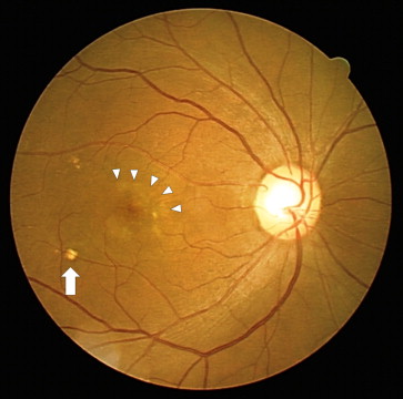
Early AMD was defined by the presence of large drusen (soft distinct and soft indistinct drusen of ≥125 μm in diameter) or RPE pigment abnormalities within the grid in the absence of late AMD in either eye. Late AMD was defined as the presence of exudative AMD or geographic atrophy (GA). Signs of exudative AMD were retinal pigment epithelial detachment or serous detachment of the sensory retina, subretinal or sub-RPE hemorrhages, and subretinal fibrous scars. GA was defined as a circular discrete area (of at least 175 μm in diameter) of retinal depigmentation with visible choroidal vessels in the absence of exudative AMD.
Information on smoking status was obtained via a self-reported questionnaire. To assess the association between the effect of cigarette smoking and the development of AMD in detail, we used 2 methods of analysis: (1) total cigarette amount using the Brinkman index, which was calculated by the daily number of cigarettes × years of smoking ; and (2) smoking status, in which the subjects were categorized as never smokers (had smoked less than 100 cigarettes in the past) and ever smokers (had smoked more than 100 cigarettes in the past).
We assessed the age- and sex-specific prevalence of early AMD and late AMD, including the phenotypes of AMD lesions. The age- and sex-adjusted standardized incidences of AMD were calculated using the direct method with reference to the World Health Organization standard population in 2010. We used analysis of variance or the χ 2 test to compare demographic characteristics. P values less than .05 were considered statistically significant.
Results
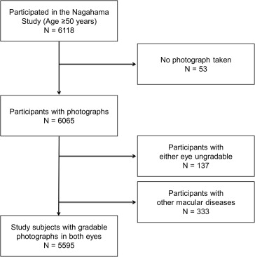
Fundus photographs were available for 6065 subjects aged ≥50 years, and 5595 of these subjects (92.3%) had photographs that were gradable for AMD lesions in both eyes ( Figures 2 and 3 ). Photographs were not taken for 53 participants because of significant media opacities, poor fixation, and/or poor participant cooperation/refusal. Photographs were ungradable in either eye (n = 137) because of media opacities (such as asteroid hyalosis of the vitreous) or poor camera focus. We excluded 333 participants with other macular disease, such as diabetic retinopathy, from the analysis. Thus, a total of 523 participants who had missing or ungradable photographs or who had macular conditions that were inadequate were excluded from this analysis. The participants with gradable photographs (n = 5595) who were included in the analyses were younger (mean age, 62.5 ± 6.5 years) than those excluded from the analysis (65.9 ± 5.9 years; P < .0001). However, no differences were found in sex between those with gradable and ungradable photographs ( P = .588). Thus, the following prevalence data are from 5595 participants with gradable photographs in both eyes.

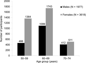
The summary of the prevalence of phenotypes of AMD in the Nagahama cohort is shown in Table 1 . In the study cohort of participants aged ≥50 years, the prevalence of soft drusen (defined as drusen of >63 μm) was 39.4% (39.2%, standardized) and that of large drusen (defined as drusen of ≥125 μm) was 17.4% (17.5%, standardized). Overall, 22.3% of all subjects had early AMD in at least 1 eye, and the prevalence increased from 16.1% in subjects aged 50-59 years to 31.2% in subjects aged ≥70 years. The overall prevalence of late AMD was 0.52% (0.58%, standardized), which increased from 0.27% in subjects aged 50-59 years to 0.98% in subjects aged 70-74 years. We found similar tendencies regarding age dependence in other features of AMD (soft drusen, large drusen, and pigment abnormalities). Whereas 10.7% of drusen subjects had pigment abnormalities, 60.4% of subjects with pigment abnormalities also had drusen. We found that subjects with larger drusen tended to have pigment abnormalities ( P < .0001). Reticular pseudodrusen were present in 38 participants (0.68%), including those that were outside of the grid; 17 cases, including a case with late AMD, were within the grid. The prevalence of reticular pseudodrusen was significantly higher among women ( P = .011), with women accounting for 32 of the 38 subjects (84.2%) with reticular pseudodrusen.
| 50-59 Years N = 1830 | 60-69 Years N = 2842 | 70-74 Years N = 923 | Total N = 5595 | Overall Standardized Prevalence a (95% CI) | |
|---|---|---|---|---|---|
| Either eye, n (%) | |||||
| Early AMD | 294 (16.1) | 665 (23.4) | 288 (31.2) | 1247 (22.3) | 22.8 (21.7-24.0) |
| Late AMD | 5 (0.27) | 15 (0.53) | 9 (0.98) | 29 (0.52) | 0.58 (0.36-0.80) |
| Soft drusen | 561 (30.7) | 1173 (41.3) | 468 (50.7) | 2202 (39.4) | 39.2 (37.9-40.5) |
| Large drusen | 216 (11.8) | 516 (18.2) | 239 (25.9) | 971 (17.4) | 17.5 (16.5-18.5) |
| Pigment abnormality | 98 (5.4) | 222 (7.8) | 71 (7.7) | 391 (7.0) | 7.6 (6.8-8.3) |
| Bilateral, n (%) | |||||
| Early AMD | 57 (3.1) | 171 (6.0) | 101 (10.9) | 329 (5.9) | 6.1 (5.5-6.8) |
| Late AMD | 0 (0.00) | 3 (0.11) | 1 (0.11) | 4 (0.07) | 0.09 (0.00-0.18) |
| Soft drusen | 61 (3.3) | 141 (5.0) | 67 (7.3) | 269 (4.8) | 4.6 (4.0-5.1) |
| Large drusen | 45 (2.5) | 127 (4.5) | 87 (9.4) | 259 (4.6) | 4.8 (4.2-5.3) |
| Pigment abnormality | 10 (0.5) | 32 (1.1) | 22 (2.4) | 64 (1.1) | 1.3 (1.0-1.7) |
a The prevalence was standardized to the World Health Organization standard population.
AMD was present in both eyes in 333 of the 1276 participants (20.7%) with any AMD. The overall prevalence of bilateral early AMD was 5.9% (6.1%, standardized), and this value increased from 3.1% in subjects aged 50-59 years to 10.9% in subjects aged ≥70 years. Bilateral late AMD was present in 4 of the 29 participants (13.8%) with any late AMD.
The prevalence of AMD according to sex is shown in Table 2 . The prevalence of early and late AMD was significantly higher in men than in women ( P = .0007 and P = .025, respectively). The subtype analysis revealed that the prevalence of RPE abnormalities was significantly higher in men than in women ( P < .0001). This tendency was found in all age groups ( P = .0001 in subjects aged 50-59 years and P = .0002 in those aged 60-69 years), although this association failed to reach significance in subjects aged ≥70 years ( P = .0694). The incidences of soft and large drusen were not significantly different according to sex ( P = .454 and P = .305, respectively).
| Male N = 1977 | Female N = 3618 | P Value | |
|---|---|---|---|
| Early AMD | 491 (24.8) | 756 (20.9) | .0007 |
| Late AMD | 16 (0.81) | 13 (0.36) | .025 |
| Soft drusen | 765 (38.7) | 1437 (39.7) | .454 |
| Large drusen | 357 (18.1) | 614 (17.0) | .305 |
| Pigment abnormality | 192 (9.7) | 199 (5.5) | <.0001 |
Finally, we evaluated the association between cigarette smoking and the development of AMD ( Table 3 ). The total amount of cigarette smoking was significantly associated with the development of early and late AMD ( P = .0153 and P = .0402, respectively). Particularly, subjects with a Brinkman index greater than 500 had a significantly higher risk for the incidence of early and late AMD ( P = .011 and P = .042, respectively, Supplemental Table 1 , available at AJO.com ). Never smokers were less likely to have early and late AMD, although these associations did not reach statistical significance ( P = .120 and P = .159, respectively). In the subgroup phenotype analysis, we found strong associations between the presence of RPE abnormalities and both the total amount ( P < .0001) and status ( P = .0003) of cigarette smoking. However, these significant associations diminished when we divided the cohort by sex ( P > .05, Supplemental Table 2 , available at AJO.com ). We found no significant association between cigarette smoking and the incidence of soft or large drusen ( P > .05).
| Brinkman Index a | Smoking Status, N (%) | |||||
|---|---|---|---|---|---|---|
| N | Mean | P Value | Ever (N = 1853) | Never (N = 3742) | P Value | |
| No AMD | 4319 | 169.9 ± 344.3 | 1405 (75.8) | 2914 (77.9) | ||
| Early AMD | 1247 | 197.2 ± 368.9 | .0153 | 435 (23.5) | 812 (21.7) | .120 |
| Late AMD | 29 | 301.9 ± 462.2 | .0402 | 13 (0.70) | 16 (0.42) | .159 |
| Soft drusen | .939 | .069 | ||||
| Absent | 3393 | 177.0 ± 348.8 | 1155 (62.3) | 2238 (59.8) | ||
| Present | 2202 | 176.2 ± 354.0 | 698 (37.7) | 1504 (40.2) | ||
| Large drusen | .305 | .798 | ||||
| Absent | 4624 | 174.5 ± 348.7 | 1528 (82.5) | 3096 (82.7) | ||
| Present | 971 | 187.2 ± 360.7 | 325 (17.5) | 646 (17.3) | ||
| Pigment abnormality | <.0001 | .0003 | ||||
| Absent | 5204 | 171.6 ± 346.4 | 1691 (91.3) | 3513 (93.9) | ||
| Present | 391 | 243.9 ± 399.9 | 162 (8.7) | 229 (6.1) | ||
a The Brinkman index was calculated by the daily number of cigarettes × years.
Results
Fundus photographs were available for 6065 subjects aged ≥50 years, and 5595 of these subjects (92.3%) had photographs that were gradable for AMD lesions in both eyes ( Figures 2 and 3 ). Photographs were not taken for 53 participants because of significant media opacities, poor fixation, and/or poor participant cooperation/refusal. Photographs were ungradable in either eye (n = 137) because of media opacities (such as asteroid hyalosis of the vitreous) or poor camera focus. We excluded 333 participants with other macular disease, such as diabetic retinopathy, from the analysis. Thus, a total of 523 participants who had missing or ungradable photographs or who had macular conditions that were inadequate were excluded from this analysis. The participants with gradable photographs (n = 5595) who were included in the analyses were younger (mean age, 62.5 ± 6.5 years) than those excluded from the analysis (65.9 ± 5.9 years; P < .0001). However, no differences were found in sex between those with gradable and ungradable photographs ( P = .588). Thus, the following prevalence data are from 5595 participants with gradable photographs in both eyes.