, Yasuya Nomura2 and Yasuya Nomura3
(1)
The Society for Promotion of International Oto-Rhino-Laryngology, Tokyo, Japan
(2)
The University of Tokyo, Tokyo, Japan
(3)
Showa University, Tokyo, Japan
Abstract
Presbycusis occurs mainly as a result of aging of the cochlea and the central auditory system. This chapter presents observations made using unconventional techniques to evaluate the nerve fibers in the organ of Corti, the basilar membrane, the stereocilia of the outer hair cells, and the stria vascularis in elderly people. Stained surface preparations of nerve fibers in the organ of Corti and the osseous spiral lamina were observed. Afferent and efferent nerve fibers were reduced in number in the lower basal turn in the elderly. Swellings of the efferent nerve fibers crossing the tunnel of Corti were occasionally observed. These swellings resembled the torpedoes seen in Purkinje cell degeneration. The medial fiber is considered an efferent fiber, and sometimes extends laterally beyond the outer hair cell area.
The stereocilia of the outer hair cells showed various types of degeneration with aging. Loss of individual hairs, fusion of several hairs, and giant cilia were common findings. After complete disappearance of the stereocilia, the surface of the cuticular plate showed remnants of equally-sized stumps, suggesting the breaking point of each hair.
In strial atrophy, the cells composing the stria vascularis disappeared with the strial capillaries. The basilar membrane of elderly people showed lipid deposits in the basal turn. These deposits were composed of neutral fat and cholesterol and were located along the filaments of the pars pectinata. As for prevention of presbycusis, an animal experiment revealed that caloric restriction prevented the age-related loss of spiral ganglion cells.
Keywords
AgingBasilar membraneCaloric restrictionCochleaNerve fiberOrgan of CortiStereociliaStrial atrophy8.1 Nerve Fibers in the Osseous Spiral Lamina
The axons of the spiral ganglion cells leave Rosenthal’s canal, and join at the central bony axis, the modiolus, to form the cochlear nerve. The dendrites of the spiral ganglion cells pass between the upper and lower shelves of the osseous spiral lamina as they head toward the organ of Corti. Near the end of the spiral lamina are small foramina called habenula perforata, through which unmyelinated nerve fibers pass to the inner and outer hair cells.
In young people, the cochlea contains abundant nerve fibers leaving Rosenthal’s canal, their bundles gradually fanning out radially in the osseous spiral lamina (Fig. 8.1). Spiral fibers intermingle with the radial fibers in the osseous spiral lamina.
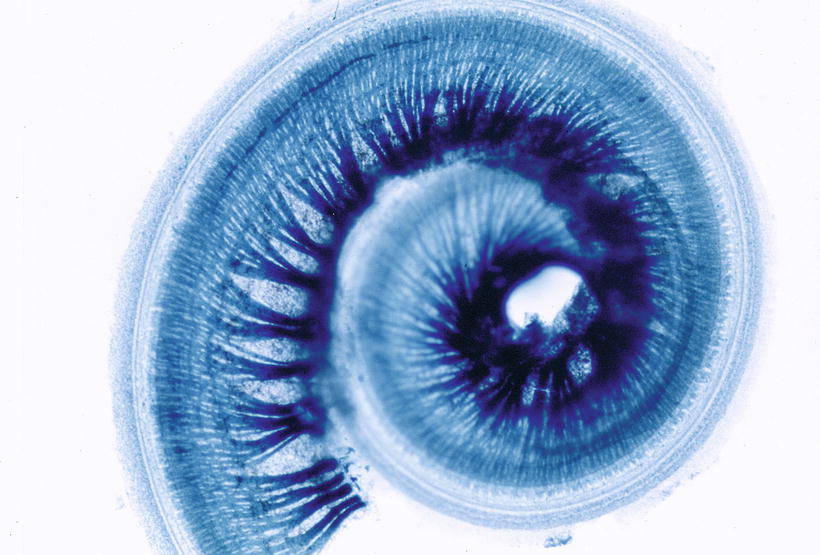
Fig. 8.1
Myelinated nerve fibers of the human cochlea. The organ of Corti is at the periphery of the specimen. Sudan Black B staining
In elderly people, the cochlea shows loss of radial and spiral fibers, particularly in the lower basal turn, with less loss toward the apex (Fig. 8.2).
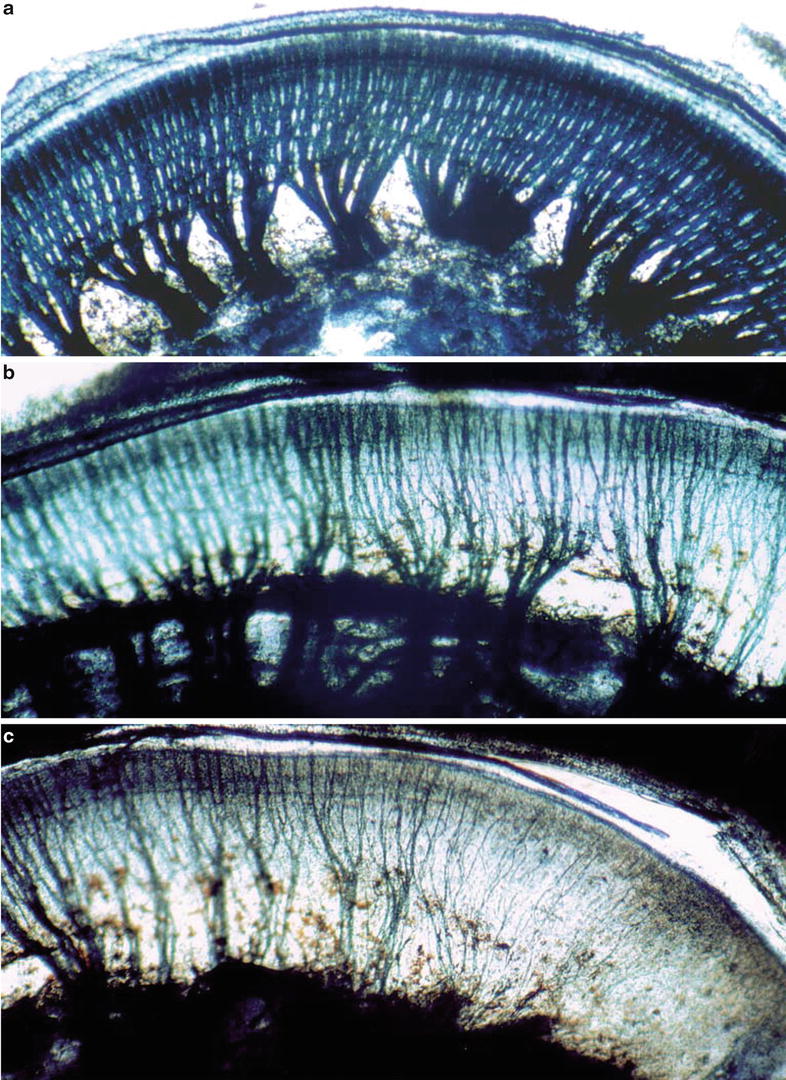
Fig. 8.2
Myelinated nerve fibers in the osseous spiral lamina. Bundles of nerve fibers emerge from Rosenthal’s canal, and fan out in the osseous spiral lamina. (a) The radial and spiral fibers are densely intermingled in the lamina of the middle turn. (b) Both radial and spiral fibers are reduced in the basal turn. (c) Almost no fibers remain near the basal end. Seventy-five-year-old woman, Sudan Black B staining, ×10
Histochemical staining for acetylcholinesterase specifically stains the efferent nerve fibers, leaving the afferent fibers unstained. From spiral bundles, fibers radiate outward, eventually terminating primarily on the outer hair cells. In the organ of Corti, the outer spiral bundle shows acetylcholinesterase activity, though postmortem autolysis may obscure details (Fig. 8.3a, b) [1, 2]. In the lower basal turn of a 59-year-old man, most of the afferent and efferent nerve fibers had disappeared, with only a few fibers remaining (Fig. 8.3c). The course of the afferent fibers was relatively simple. By contrast, the efferent fibers followed a complicated course (Fig. 8.4). No efferent fibers were seen in the osseous spiral lamina of the cochlea (Fig. 8.5). Clinical data were not available for this individual.
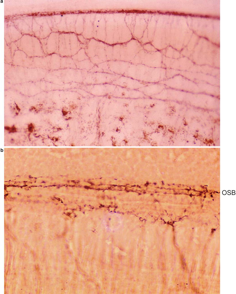
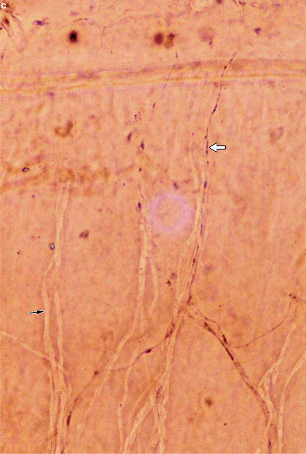
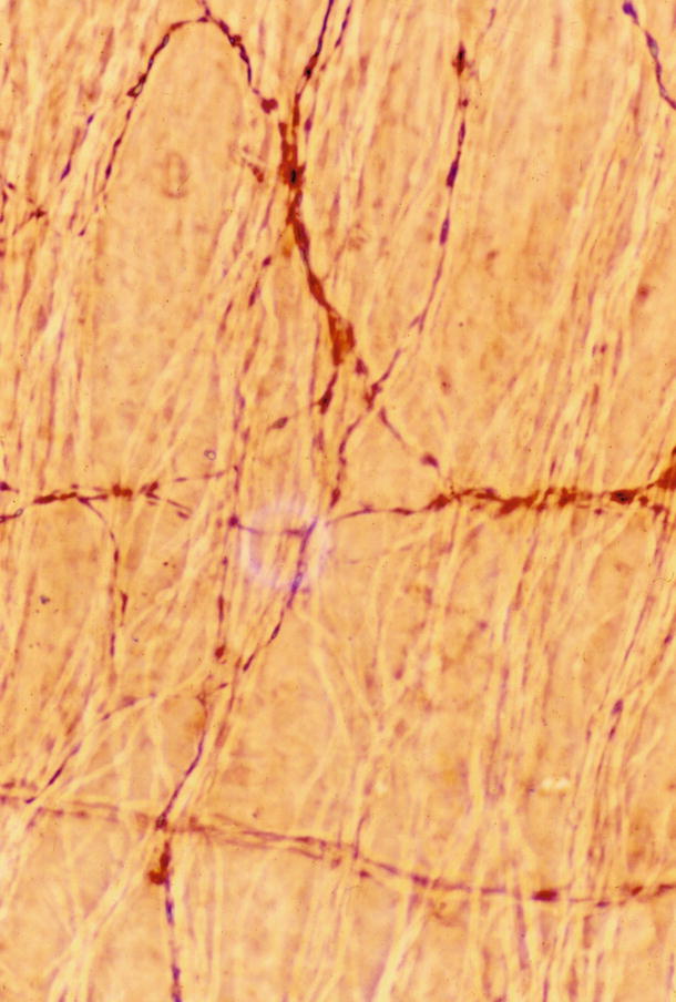
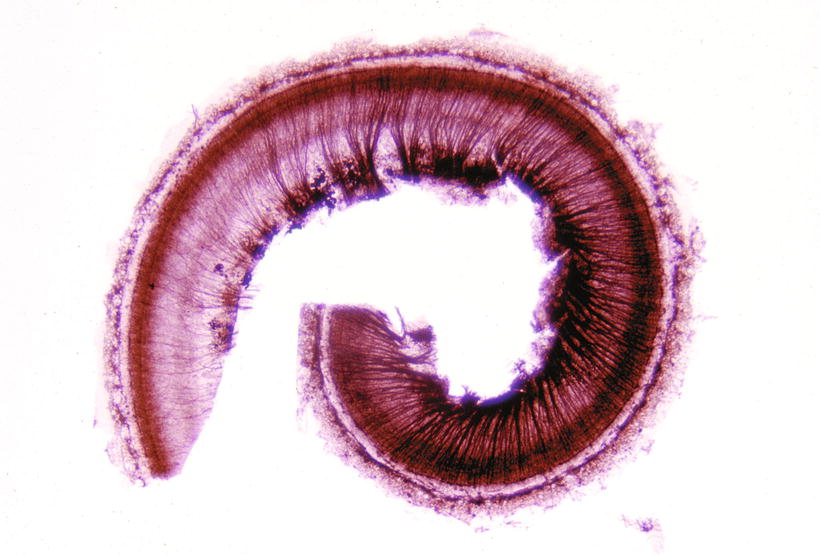
Fig. 8.3
Distribution of efferent fibers in the organ of Corti and osseous spiral lamina. (a) Strong staining at the top of the photograph indicates acetylcholinesterase activity in the organ of Corti (×45). (b) High power view of the organ of Corti. Sparse enzyme activity due to marked loss of the organ of Corti and postmortem changes. OSB outer spiral bundles (×160). (c) A single efferent fiber (white arrow) and a few afferent fibers (black arrow) remain in the lower basal turn (×160). Fifty-nine-year-old man, staining for acetylcholinesterase
Fig. 8.4
Efferent and afferent fibers in the osseous spiral lamina. Because of the difficulty of pursuing the course of an individual fiber, an area with reduced fibers is selected for observation. Generally, the afferent fibers run radially, whereas the efferent fibers run spirally with a complicated course before taking a radial course to the organ of Corti. unstained fiber afferent fiber, stained fiber efferent fiber ×160
Fig. 8.5
The cochlea shows reduced numbers of afferent fibers, particularly in the basal turn, and complete loss of the efferent fibers. Seventy-eight-year-old man with severe hearing loss, OTAN staining
Gacek [3] reported a case in which the cochlea showed only efferent fibers.
8.2 Nerve Fibers in the Organ of Corti
It is difficult to follow nerve fibers in the organ of Corti in serial temporal bone specimens. However, they are easier to follow in properly stained surface specimens of the cochlea. When observed as a surface preparation, the tunnel of Corti is seen as a wide, clear belt because of its sparse cellularity. Individual nerve fibers can be clearly observed here (Figs. 8.6 and 8.7). The medial fiber and basilar fiber cross the tunnel of Corti. The medial fiber crosses the medial part of the tunnel, and is the efferent fiber [4]. The basilar fiber crosses the tunnel at its base and is the afferent fiber (Fig. 1.19).
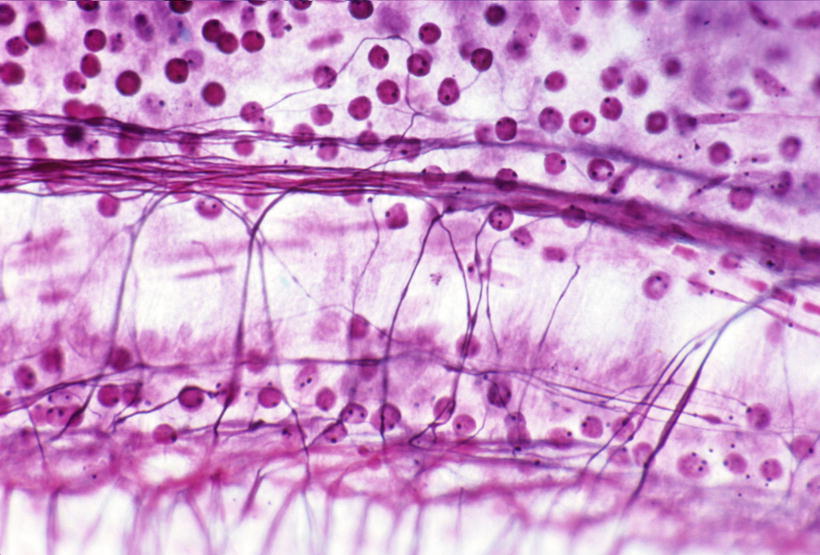
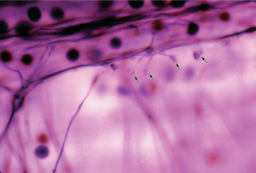
Fig. 8.6
Nerve fibers in the organ of Corti of the apical turn. There are fewer fibers near the apical end. Holmes staining (original ×40)
Fig. 8.7
Nerve endings (arrows) at the first row of the outer hair cells. Holmes staining (original ×100)
According to Spoendlin [5], 90 % of the afferent fibers in the cat terminate on the inner hair cells, while 10 % terminate on the outer hair cells.
A similar finding was observed in a human organ of Corti, where only the basilar fiber was observed in the tunnel of Corti in a 6-month-old fetus after Holmes staining. The basilar fibers ran at the base of the tunnel, maintaining regular spacing. Evaluation revealed that 10–15 % of the nerve fibers crossed the tunnel to reach the outer hair cells after passing through the habenula perforata (Fig. 8.8) [6].
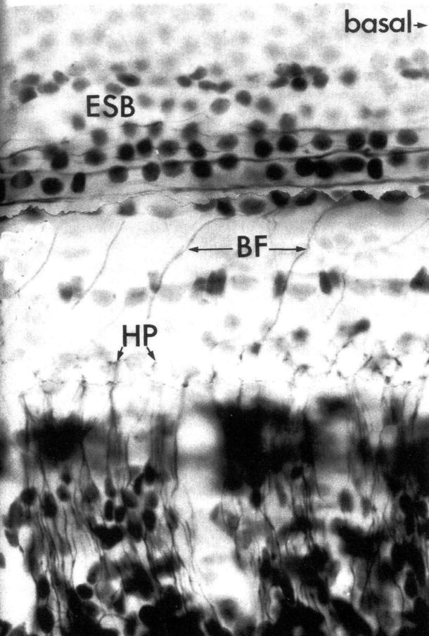
Fig. 8.8
Basilar fibers crossing the tunnel, maintaining a regular interval. No medial fibers are seen [6]. ESB external (outer) spiral bundle, BF basilar fiber, HP habenula perforata. The basal direction is to the right in the figure. Six-month-old fetus, Holmes staining
Some efferent fibers have the appearance of a gigantic old tree. Perhaps the composing fibers loosen in the process of degeneration (Fig. 8.9).
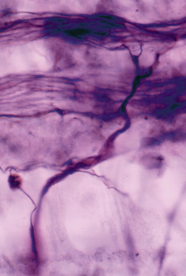
Fig. 8.9
Gigantic efferent fibers join the outer spiral bundles. Possibly in the process of degeneration. Eighty-four-year-old woman, Holmes staining (original ×100)
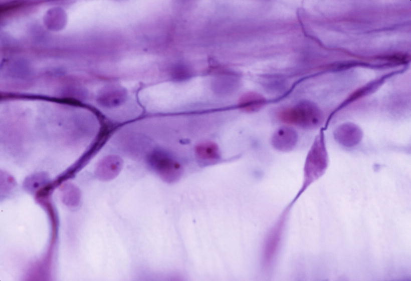
A striking finding in the medial fibers is the presence of spindle-shaped swellings, similar in appearance to torpedoes (Figs. 8.10–8.12). These fibers are generally thick, and after forming a torpedo shape they bifurcate before joining the outer spiral bundles.
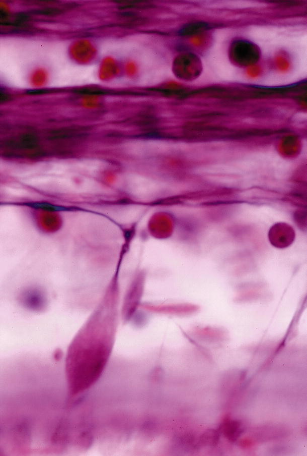
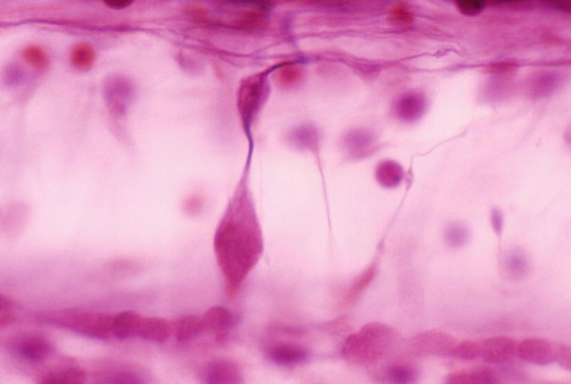
Fig. 8.10
Axonal swelling of an efferent fiber creating a torpedo. Maximal diameter is about 8 μm. Many torpedoes were present in this specimen. The patient had no neurological disease. Forty-six-year-old woman, Holmes staining (original ×100)
Fig. 8.11
Two torpedoes connected in the same nerve fiber. Forty-six-year-old woman (original ×100)
The spindle-shaped swelling observed in the efferent fibers appears similar to Purkinje cell axonal torpedoes. According to Louis et al. [7], a prominent finding in the cerebellum of patients with essential tremor is an abundance of torpedoes, swellings that result from the mis-accumulation of cell constituents. Immunoreactivity for phosphorylated neurofilament protein revealed clear labeling of torpedoes. On electron microscopy, torpedoes were packed with randomly arranged 10–12 nm neurofilaments. Mitochondria and smooth endoplasmic reticulum were abundant at the periphery of the torpedoes. The authors concluded that the torpedoes in essential tremor represent the mis-accumulation of disorganized neurofilaments and other organelles.
Efferent nerve fibers run spirally in the osseous spiral lamina toward the apex. In the cochlea of elderly people, both afferent and efferent fibers show the greatest loss in the lower basal turn. The torpedoes were rarely observed in aged cochleae, whereas they sometimes appear in younger individuals, where they are evenly distributed throughout the cochlea. The torpedoes might suggest a degenerative process of the nuclei of the efferent fibers.