Postoperative Glaucoma
M. Edward Wilson
Rupal H. Trivedi
John M. Facciani
William J. Johnson
Postoperative glaucoma after childhood cataract surgery remains a major concern despite all of the improvements in technology and the increased use of intraocular lenses (IOLs). To date, scientists cannot submit an etiology that is accepted by all clinicians, clinicians cannot offer a treatment that is effective for all patients, and the onset of glaucoma can range from the immediate postoperative period to many years later. To make matters worse, children do not always cooperate for intraocular pressure (IOP) measurements, optic disc evaluation, or visual field documentations. Thus, defining when glaucoma is present and when a treatment is effective at halting the progression of damage is unusually challenging. The common factor for all of these patients is the fact that cataract surgery has been performed. Even with severe microphthalmia, glaucoma does not seem to occur in the absence of the cataract procedure. In the predisposed eye, surgery to remove the cataract may trigger a cascade of events that can lead to elevated IOP and/or glaucoma early on or even 5 to 15 years later. Ophthalmologists must be vigilant about assessing for postoperative glaucoma even after uncomplicated cataract removal in young children.
How often does late postoperative glaucoma occur? Why does it occur? Which patients are more likely to develop glaucoma after surgery? What therapy is appropriate once this is recognized? These questions are discussed in this chapter.
ESTIMATE OF FREQUENCY OF POSTCATARACT GLAUCOMA IN CHILDREN
The incidence of glaucoma following pediatric cataract removal has been reported to be as low as 5% and as high as 41%.1,2,3,4,5,6 In a review of 13 studies from the international literature on congenital cataract surgery in an earlier era, Francois3 reported that delayed glaucoma following cataract surgery in children occurred from 0% to 14% of cases. The majority of these data are available in reports from the 1940s or 1950s, when the linear aspiration technique was used. This older method resulted in lens swelling and, at times, a prolonged flat anterior chamber. Acute angle-closure glaucoma and excessive inflammation often ensued. Scheie7 introduced an aspiration technique in 1960 that may have reduced the incidence of complications compared to the linear extraction method. In 1984, Chrousos et al.2 reported their 15-year experience with pediatric cataract surgery; the surgeons utilized Scheie’s manual aspiration technique as well as automated styles of cataract removal. The standard needle-and-syringe technique was performed in 304 eyes, rotoextraction with a small opening in the posterior capsule was performed in 34 eyes, and Ocutome vitrector aspiration with wide posterior capsular excision was performed in 54 eyes. All patients had at least 3 months of follow-up. Overall, chronic glaucoma was found in 6.1% of the eyes; no patients with Ocutome aspiration developed glaucoma. Interestingly, the 6.1% overall rate of glaucoma reported by Chrousos et al.2 was similar to the 5% rate achieved by Francois3 before aspiration techniques were used. The authors of the 1984 study admitted that a limitation of the study is the length of follow-up of some patients, which could have resulted in underreporting glaucoma. While the aspiration-only group was followed for a mean of 6.3 years, the Ocutome group was followed for only 2 years on average.
Making comparison between studies is rather unreasonable for many reasons, including the variety of definitions of glaucoma that authors have used through the ages and the variety of instruments used to measure IOP. However, perhaps the most important factor in studies aiming to report the frequency of glaucoma in a postoperative population is the length of follow-up, as a study by Simon and associates addresses below.
Simon et al.5 invited their patients who had undergone pediatric cataract surgery back to their office to check for asymptomatic glaucoma, which was defined as an IOP ≥26. While almost a quarter of the patients examined had glaucoma, the interesting data involved length of follow-up. While only 1 of 14 eyes (7%) with ≤60 months of
follow-up since lensectomy developed glaucoma, 7 of 17 eyes (41%) followed for >5 years had glaucoma. This suggests that reports with <5 years of follow-up may significantly underestimate the frequency with which glaucoma develops after pediatric cataract surgery. Chrousos et al.2 and Simon et al.5 both pointed out an important finding in asymptomatic postoperative glaucoma—that although the onset is typically delayed until years after the surgery, the diagnosis may be made within the first year of surgery in some cases and surveillance of IOP must be initiated early.
follow-up since lensectomy developed glaucoma, 7 of 17 eyes (41%) followed for >5 years had glaucoma. This suggests that reports with <5 years of follow-up may significantly underestimate the frequency with which glaucoma develops after pediatric cataract surgery. Chrousos et al.2 and Simon et al.5 both pointed out an important finding in asymptomatic postoperative glaucoma—that although the onset is typically delayed until years after the surgery, the diagnosis may be made within the first year of surgery in some cases and surveillance of IOP must be initiated early.
Simon et al.5 did not choose objective signs of glaucoma such as visual field loss or optic nerve head damage in their definition of glaucoma. Therefore, it is likely that some of his patients actually had ocular hypertension (OHTN), and “true” glaucoma probably occurred less frequently than was reported. In adults, the Ocular Hypertension Treatment Study (OHTS)8 recognized that only a small percentage of patients with an IOP >21 mm Hg will progress to develop glaucoma after several years.
A prospective, nonrandomized study provides some valuable data on the rates of pediatric glaucoma and OHTN after automated lensectomy and vitrectomy. Egbert et al.9 reviewed records of patients with 5 years of follow-up after lensectomy. Of 159 patients, 52 were excluded because of trauma, microphthalmos, uveitis, or a similar complicating factor. Sixty-two of 107 patients (58%) participated, and participating and nonparticipating patients had no differences in average corneal diameter or age at the time of surgery. Glaucoma was defined as an IOP >21 with a cup/disk ratio >0.5 or a cup/disk asymmetry ≥0.2. OHTN was defined as an IOP >21 without the aforementioned optic nerve parameters. Six of 40 (15%) patients had glaucoma in their aphakic eye. OHTN was even more common. Thirteen of forty (32.5%) patients had OHTN in their unilaterally aphakic eye, while 10 of 22 (45%) patients who previously had bilateral cataracts had OHTN in one (four patients) or both (six patients) eyes. Overall, 23 of 62 (37%) patients had aphakic OHTN when examined ≥5 years after surgery. This report demonstrates that OHTN is a common long-term finding in patients with pediatric aphakia and glaucoma is less common but not rare event. With time, more patients with a normal IOP or with OHTN may develop glaucoma.
In an additional study by Egbert et al.,10 OHTN and glaucoma rates were examined for patients 5 and 10 years after undergoing automated lensectomy and vitrectomy. Their data showed a significant trend of glaucoma development with additional years after surgery. They showed a 23% conversion rate from OHTN to glaucoma, with similar definitions for the respective findings as their earlier paper. This occurred during a mean observational period of 7.2 years. Also shown were the rates of glaucoma in the 63-patient cohort at 5 (7/63; 11%) and 10 years (5/59 remaining; 9%) after surgery. These findings suggest that the risk of aphakic patients developing glaucoma does not dramatically change in the first 10 years postoperatively, demonstrating the necessity of continued follow-up. So far, evidence has not been offered to suggest that the risk of glaucoma in aphakic eyes ever subsides; thus follow-up should be pursued indefinitely. Swamy et al.11 also noted the lifelong risk of developing secondary glaucoma in their 2007 review of 423 eyes over 20 years that received cataract removal with or without primary IOL. Their data demonstrated a range of time to glaucoma onset postoperatively of 2 weeks to 16.8 years. These two studies by Egbert and one by Swamy demonstrate the necessity of continual follow-up with patients after surgery.
We believe that it is vital to examine patients on at least a yearly basis after uneventful pediatric cataract surgery with or without IOL implantation. Egbert et al.9 concluded that good data can be obtained from patients >5 years of age in the clinic. We schedule examinations under anesthesia (EUA) whenever clinic examinations with IOP measurements, refractions, serial axial length measurements, and optic nerve evaluations cannot be done awake in the clinic. Baseline optic disc documentation should be performed (Fig. 51.1). Early in childhood, EUA is often done yearly, but by age 5 to 6, the examination in the office is more reliable (except in those children with
developmental delay). As discussed below, the Icare (Icare USA, Raleigh, NC) rebound tonometer has reduced the need for EUA in some children due to its ease of use and the fact that no topical anesthetic needs to be instilled.
developmental delay). As discussed below, the Icare (Icare USA, Raleigh, NC) rebound tonometer has reduced the need for EUA in some children due to its ease of use and the fact that no topical anesthetic needs to be instilled.
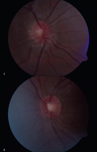 Figure 51.1. Disc photo of 11-year-old pseudophakic child diagnosed as glaucoma suspect due to high IOP. |
EXAMINATION UNDER ANESTHESIA
When evaluating under anesthesia, the method of anesthetizing must be considered due to the interaction between anesthetic agents and IOP. In the literature, there are many resources describing the effect of anesthesia methods on IOP. In their book series, The Glaucomas, Sampaolesi et al.12 provided an effective overview of the impact of different medications potentially used during an EUA. They reviewed the effects of the more historical agent, ether, with its increase in IOP from the Valsalva-like response, and then discussed differences among more modern agents including barbiturates, fluorinated inhalation agents, etc. Though all general anesthetic agents affect IOP, Sampaolesi and associates point out that the biometric measurement of axial length is not affected by IOP fluctuations. This allows axial length to be assessed as another interpretable finding independent of anesthesia for evaluating glaucoma beyond just an IOP measurement. As outlined by Sampaolesi and associates, barbiturate dosage is difficult to accomplish accurately. Too little and the patient begins to regain consciousness and struggle, raising IOP, and too much will decrease IOP due to blood pressure decrease from respiratory depression. Conversely, if succinylcholine is used to paralyze respiratory movements, the blood pressure spike facilitates an increased IOP. The inhalation agents halothane (Fluothane), methoxyflurane (Penthrane), and sevoflurane (Sevorane) categorically lower IOP. However, halothane and methoxyflurane represent older generation agents largely eclipsed by sevoflurane in modern settings. In addition to practical advantages such as being well tolerated and having a quick recovery, sevoflurane maintains the effect of its predecessor halothane in maintaining the eyes in a straight ahead position while deeply anesthetized, helping facilitate accurate assessment of clinical parameters during an EUA. In a setting with controlled ventilation and normocapnia, sevoflurane causes a proportional decrease in IOP compared to depth of anesthesia. A proportional relationship offers the ability to interpret the IOP figures for a nonanesthetized state when evaluating for glaucoma.
Nagdeve et al. demonstrated that ketamine in a low dose (3 mg/kg) does not affect IOP. This was shown to contrast with an induction dose of ketamine, which raised IOP after administration.13 Additionally, Jones et al.14 demonstrated the effect of sequential treatment of pediatric patients with ketamine and sevoflurane. They showed that IOP measurements while anesthesia was maintained using sevoflurane were significantly lower than measurements taken after ketamine was administered. This effect of sevoflurane remains consistent with the effect of an earlier inhalation agent, halothane. While the Jones study conceded their small, retrospective sample size (16 eyes from 8 patients) was a limitation, components from their study are demonstrated independently elsewhere. These studies taken together offer the opportunity to use a non-induction dose of a dissociative anesthetic, ketamine, for collecting accurate IOP measurements, with sevoflurane utilized for the balance of the EUA, offering its many advantages over ketamine for anesthesia.
Recently, a new instrument has become available for the evaluation of IOP, the Icare rebound tonometer (Icare TAO1i) (Icare USA, Raleigh, NC). This instrument allows for IOP evaluation without the need for a topical anesthetic. In a study by Lundvall et al.15 in 2011, 46 infants were recruited into this evaluation of the Icare tonometer while awake in the clinic. Only seven refused to cooperate. The remaining 39 were evaluated successfully. This new technology allows for the possibility of avoiding the operating room for EUAs, if the only indication is an inability to acquire an accurate IOP measurement. Even if the operating room cannot be avoided for other parts of an EUA, the Icare allows for more frequent evaluation of IOP outside the operating room in the infant population. Until recently, the Icare was limited by its mechanism requiring the patient to be upright to allow for proper measurement; however, the latest model (Icare Pro) allows for supine patients, providing additional flexibility.
Central Corneal Thickness
Central corneal thickness (CCT) has been more recently studied with respect to pediatric cataract surgery patients. Goldmann applanation tonometry has been the gold standard for IOP measurement, but variability in CCT affects applanation tonometry measurement. Simsek et al.16 published a study in 2006 outlining the involvement of CCT in pediatric cataract surgery. They noted CCT of aphakic and pseudophakic patients to be significantly higher than age-matched controls. Muir et al.17 compared the CCT of normal children (controls) and in those with cataract and those operated for cataract. In the absence of factors known to affect CCT (Down syndrome, Marfan syndrome, and aniridia), CCT is similar in eyes with pediatric cataracts and normal control and increases after cataract surgery. Further data from Muir lend corroboration to this theory. Unoperated unilateral cataracts (n = 9) in their study were shown to correlate strongly (r2 = 0.7) with a nonzero slope (P = 0.005) to the CCT of the fellow noncataractous eye. Time after surgery is shown to be a positive predictor for CCT. The CCT of aphakic eyes without glaucoma in this study were assessed against postoperative years. The slope offered a statistically nonzero P-value (<0.001) and a positive correlation
between increasing CCT and years after lensectomy (r2 = 0.6). The data analyzed for these results even included developmental, persistent fetal vasculature (PFV), and microphthalmic cataracts because their CCT values were not significantly different from the congenital cataract CCT cohort. A difference was noticed in the comparison of aphakic eyes with glaucoma and those aphakic eyes without glaucoma, although the authors postulated that perhaps there was an unintentional selection bias in those eyes because of a predisposition for thicker corneas and glaucoma or that the glaucoma contributed to increased CCT via subclinical edema.
between increasing CCT and years after lensectomy (r2 = 0.6). The data analyzed for these results even included developmental, persistent fetal vasculature (PFV), and microphthalmic cataracts because their CCT values were not significantly different from the congenital cataract CCT cohort. A difference was noticed in the comparison of aphakic eyes with glaucoma and those aphakic eyes without glaucoma, although the authors postulated that perhaps there was an unintentional selection bias in those eyes because of a predisposition for thicker corneas and glaucoma or that the glaucoma contributed to increased CCT via subclinical edema.
In addition to the data comparisons, Muir et al.17 noted a potential disparity between the findings of CCT in postoperative cataract children and the CCT findings detailed in the OHTS. OHTS work yielded a model describing increasing CCT as a protective measure against glaucoma development. Conversely, this study demonstrated that CCT among aphakic eyes with glaucoma was further increased beyond the typical nonglaucoma postoperative increase in CCT. Muir pointed out the potential inconsistency in applying the OHTS results from adults to aphakic children. If increasing CCT protects against glaucoma, why does a significant minority of patients develop glaucoma after surgery to remove a cataractous lens? These findings highlight a potential dichotomy in the interpretation of CCT changes in aphakic pediatric eyes compared with natural adult eyes. Endothelial corneal damage or an inherent response to aphakia/pseudophakia was offered as potential explanations of this finding of increased CCT postoperatively. Further, for those patients developing glaucoma postoperatively, it was “tempting” for the authors to interpret the additional CCT increase as the result of increased IOP, leading to added corneal endothelial injury and subclinical stromal edema.
PROPOSED MECHANISMS FOR THE DEVELOPMENT OF GLAUCOMA AFTER PEDIATRIC CATARACT SURGERY
The causes of pediatric aphakic open-angle glaucoma are as elusive today as they were years ago. In 1977, Phelps and Arafat18 brought this to the attention of ophthalmologists as a “warning.” They were surprised to diagnose an insidious, asymptomatic type of glaucoma in 18 patients who had undergone congenital cataract removal years (6-56 years) before the patients manifested an elevated IOP. They expressed concern that although those patients had cataracts removed by simple needling, linear extraction, or intracapsular extraction, more modern techniques (such as needling and aspiration, phacoemulsification, and rotoextraction) may also produce glaucoma.
The authors discussed several of the mechanisms that could be at fault. Could an undescribed ocular syndrome involving both cataract and glaucoma be causal? Does early surgery promote such significant inflammation or expose the fledgling trabeculum to so much lens protein that it is irrevocably damaged? Is a vitreous component toxic to the trabeculum? Even today we do not know the answer to these questions or how much these factors contribute to the development of glaucoma, if at all.
Late-onset open-angle glaucoma occurs much more commonly than angle closure after pediatric cataract surgery.1,4,5,6 Of the 11 eyes with glaucoma for which gonioscopic data were available, all had open angles.2 Open-angle glaucoma can occur without the presence of any symptoms or gross changes in the appearance of the eye and has been described both in normal-appearing open angles and in angles that have undergone change. Acute angle closure following pediatric cataract surgery with modern techniques is relatively uncommon, and peripheral iridectomies are performed less commonly today than in years past.1,4,5,6 Kang et al.19 discussed their findings of a bimodal distribution. Early onset was found to favor angle closure, while late onset was found to occur more frequently in an open-angle fashion. In 1986, Walton20 discussed pupillary block and chronic angle closure from peripheral anterior synechia as the typical mechanism following cataract removal by the “aspiration” mechanism. A decade later, Walton’s American Ophthalmological Society thesis6 concluded that the asymptomatic, postoperative glaucoma in aphakic patients was actually an open-angle mechanism. Walton6 studied the angle structure of 65 aphakic children with glaucoma. Vitrectomy techniques were utilized in the majority (80%) of cases. Preoperatively, the majority of patients with available gonioscopy (19/29 eyes) had no angle abnormalities, while 10 patients did have “anomalous attachments from the iris root to Schwalbe line and the trabecular meshwork.” Postoperatively, the angles were open in 79 of 80 eyes, but in 76 of 79 (96%) eyes, “circumferential repositioning of the iris insertion anteriorly at the level of the posterior or mid-trabecular meshwork with resultant loss to view of the ciliary body band and scleral spur” occurred. Windows of visible scleral spur or ciliary body were noted in these eyes, confirming open angles. Phelps and Arafat18 had described similar changes in the anterior chamber angles in their patients. Walton6 observed scattered pigment deposits in the exposed anterior trabecular meshwork and, less frequently, white crystalline deposits suggestive of lens protein.
The mechanism does not appear to be related to clinically identifiable late postoperative inflammation. Walton6 reported on slit-lamp examinations of 19 children performed after lensectomy and before glaucoma was diagnosed. In none of those patients was an anterior chamber cell or band keratopathy present; these patients had a
different appearance than those with chronic inflammation. In addition, none of the patients examined after the diagnosis of glaucoma had evidence of active intraocular inflammation.
different appearance than those with chronic inflammation. In addition, none of the patients examined after the diagnosis of glaucoma had evidence of active intraocular inflammation.
The absence of active intraocular inflammation years after surgery does not negate the possibility of acute postoperative inflammation causing significant, immediate damage to the trabecular meshwork that develops into a chronic, secondary open-angle glaucoma. Several authors have reported cases of bilateral cataracts that had subtle, bilateral angle abnormalities but developed glaucoma only in the operated eye.6,21 We are curious whether the presence of “anomalous attachments from the iris root to Schwalbe line and the trabecular meshwork” documented in 10 of Walton’s6 patients (but absent in 19 patients) before surgery implies that an abnormal process was already occurring subclinically in all eyes. Perhaps cataract surgery simply amplified the process such that “circumferential repositioning of the iris insertion anteriorly” was observed at a later time. Phelps and Arafat18 observing similar gonioscopic findings in patients after surgery implied that the uniformity of the angle findings “throughout its circumference” instead suggested that these findings were congenital and not related to the cataract surgery. There is no way to prove, however, that those angle findings were not indicative of subclinical dysfunction.
Walton6 himself, in the discussion following his study, lamented that despite his careful attention to gonioscopic detail in his patients, the cause of the glaucoma could not be inferred. Whether or not the angle is described as open or closed is probably irrelevant if we cannot correlate microscopic, ultrastructural changes (by light microscopy or electron microscopy) in the trabecular meshwork with changes in aqueous outflow and increases in IOP in eyes of children with and without glaucoma who had cataract surgery in one or both eyes. Such a study may lead to the elucidation of the mechanism of the glaucoma and, ultimately, its true cause.
It is possible that cataract extraction may indeed damage a growing, vulnerable (“immature” or “developmentally arrested”) anterior chamber angle in an eye with a subclinically imperfect trabecular outflow in a way that creates high IOP years later.4 This may be why patients with a preexisting ocular abnormality (such as trauma, dislocated lens, chronic uveitis, or anterior segment dysgenesis) may be at higher risk for postoperative glaucoma.6
If such a theory were correct, and if surgery were to be performed satisfactorily with modern techniques, the most important step in preventing glaucoma for these patients would be to minimize acute postoperative inflammation by applying various forms of anti-inflammatory preoperative and postoperative medications. The results of a randomized, prospective trial addressing this issue would be most helpful. Until then, we operate on eyes with significant cataracts using the least traumatic techniques available and hope we are doing more good than harm. Then we treat glaucoma as we detect it.
RISK FACTORS HISTORICALLY ASSOCIATED WITH POSTCATARACT, PEDIATRIC GLAUCOMA
Clinicians can care for their patients better by identifying those who appear to be at risk for developing the disorder. A number of reports have discussed the following risk factors associated with postoperative glaucoma in children with cataracts: microcornea, poorly dilating pupils, surgery at <1 year of age, the presence of other ocular disease (e.g., congenital rubella syndrome), nuclear cataract, PFV, and performance of a posterior capsulorhexis. Much disagreement accompanies these risk factors, and some authors found no association among glaucoma, age at surgery, microphthalmos, and surgical complications.5
Age at Surgery
Mills and Robb4 reported risk factors for childhood glaucoma: cataract surgery at an age of <1 year (relative risk [RR] = 9.9; P ≤ 0.001), microcornea (RR = 4.4; P ≤ 0.001), poor pupillary dilation (RR = 5.2; P ≤ 0.001), and congenital rubella syndrome (RR = 5.8; P ≤ 0.001). The RR was notably high for patients undergoing surgery before the age of 6 months (RR = 5.4; P ≤ 0.001) and 1 year (9.9; P ≤ 0.001). No patient who had surgery after 1.25 years of age developed chronic open- or closed-angle glaucoma. The authors state,
The time at surgery may not be independent of other pathologic factors… [as] a disproportionate share of those patients who had early cataract surgery had other ocular abnormalities (congenital rubella syndrome (10.1% of 79 eyes operated on before 1 year of age), poorly dilating pupils (22.0%), microcornea (10.1%), or persistent fetal vasculature (6.3%)) … or more complete lens opacity.
The majority of Walton’s patients (77%) were also operated on at <1 year of age. The author suggests that performing surgery on “small eyes with small corneas and often poorly dilating pupils must be considered a risk factor for the development of glaucoma.” The author implies that cataract surgery is difficult to perform adequately on these eyes and cites residual lens tissue behind the iris in 78% of patients and a prominent need for secondary lens surgery (in 47% of patients) as evidence of technical inadequacy of the surgery in these patients. Walton6 argues that the angle abnormalities are the result of cataract surgery—especially early surgery. The immature trabecular meshwork of patients undergoing cataract surgery at a very young age was exposed to inflammation or direct surgical trauma and led to glaucoma.
Magnusson et al.22 prospectively followed a cohort of 137 patients in Sweden for an average of 9 years and concluded that cataract extraction in children <10 days old is associated with double the frequency of glaucoma. Twenty-nine percent (4/14) of patients operated on before the age of 10 days developed glaucoma; operations performed after 10 days of life had half the frequency of glaucoma. A 2004 article by Peter Rabiah23 has made it more difficult to retain the belief that age at surgery is unrelated to the development of glaucoma. Five hundred seventy eyes of 322 patients who underwent limbal approach surgery without IOL implantation in Saudi Arabia were analyzed. Patients were excluded if follow-up was for <5 years or if ocular trauma, PFV, prior eye surgery, or rubella or Lowe syndrome was present. Microcornea was not an exclusion criterion. Glaucoma was defined as an IOP ≥26 mm Hg on two occasions. Potential predictors of risk were entered into a univariate and multivariate model. Glaucoma was diagnosed in 118 of 570 eyes (21%) at a mean age of 5.4 years after surgery (range 2 weeks-15.6 years); average total follow-up for eyes with and without glaucoma was 8.5 and 10.9 years, respectively. The vast majority (86%) of the glaucoma was diagnosed in patients who underwent surgery at or before 9 months of age. Of patients with cataracts in one or both eyes, no unoperated fellow eye developed glaucoma. The significant predictors of glaucoma in the multivariable analysis included microcornea, primary posterior capsulotomy/anterior vitrectomy, secondary membrane surgery, and surgery at ≤9 months of age. The risk appeared substantially lower in children operated on after 3 years of age. The significantly lower rate of glaucoma with late surgery in these patients lends credence to early age at cataract surgery as an additional, independent risk factor for postsurgical glaucoma.
Chen et al.24 offered similar conclusions in their 2006 article. They retrospectively reviewed pediatric glaucoma patients with previous cataract surgery in their center from 1970 to 2003. This produced a cohort of 368 eyes of 258 patients to analyze for aphakic glaucoma risk factors. Two hundred and sixteen eyes (58.7%) of 150 patients in this group developed aphakic glaucoma. They assessed significant risk factors, and the most prominent (P < 0.001) were cataract removal before 1 year of age and postoperative complications. We noted that all eyes in which glaucoma developed had cataract surgery before 4.5 months of age when eyes with known glaucoma risk factors were excluded.25 In addition, all eyes that developed glaucoma had cataract diagnosed within the first month of life. Michaelides et al.26 also noted the significance of age as an aphakic glaucoma risk factor. In their review of 71 eyes with at least 5 years of follow-up, the average age at surgery for the eyes developing glaucoma was 1.6 months compared to 28.7 months old at surgery on average for the eyes not developing aphakic glaucoma. Among patients who undergo surgery before a year of age (specifically before 10 months of age in this study), Khan and Al-Dahmash27 noted the lowest risk for developing aphakic glaucoma when surgery was performed between 3 and 4 months of age. The authors cautioned against waiting much beyond about 4 to 6 weeks of life to remove infantile cataracts due to the amblyopia risk. Wong et al.28


Stay updated, free articles. Join our Telegram channel
Full access? Get Clinical Tree
