Chapter 34
PHOTIC AND ELECTRICAL TRAUMA

It has long been recognized that abnormally high amounts of light can damage the retina (gazing at the sun resulted in visual loss). In addition, today there are artificial light sources capable of causing photic retinal injury (e.g., ophthalmic instruments produce increasingly intense light to meet the demands of ophthalmologists for better diagnostic and therapeutic tools). The occupational use of laser systems is growing, and there is grave concern about the application of lasers as weapons specifically targeted at the human eye.
In this chapter we outline the basic concepts concerning the interaction of light with the eye and describe the clinical course of different types of light-induced eye injuries. We also provide guidelines for prevention, which—in the absence of any effective treatment for retinal phototoxicity—currently appears to be the only realistic approach at our disposal. Electrical ocular injury is also discussed.
PHOTIC TRAUMA
Pathophysiology
Light radiation in the visible and near IR part of the electromagnetic spectrum enters the eye and is focused by the refractive media to a retinal image about 5-30 µm in diameter. Focusing increases the retinal irradiance (see Table 34–1) by a factor of well over 10,000 above the irradiance incident at the cornea.
| Energy | The capacity for doing work, measured in J. Energy is commonly used to express the output of pulsed lasers such as Nd:YAG | |
| Power | The rate of energy delivery, expressed in watts (W; joules per second). Power is used to express the output of continuous-wave lasers such argon lasers | |
| Irradiance | Radiant power per unit area incident upon a given surface, such as the retina, measured in watts per unit area (e.g., W/cm2) |
PEARL… The nature and severity of photic damage depend on several factors related to the light source and the eye.
Source-Related Factors
Wavelength of the Radiation Optical radiation (wavelength: 380–1400 nm) is transmitted and focused on the retina by the eye’s media.1,2 Retinal exposure to such radiation at an above-threshold dose will cause damage. Photons of shorter (UV) or longer (far IR) wavelengths are rapidly absorbed in organic tissues and may cause corneal injury.
Duration and Intensity of Exposure Three distinct mechanisms of light-induced tissue injury have been described, although more than one might be responsible for damage under certain circumstances.
• Thermal damage occurs when excessive energy (see Table 34–1) is absorbed by a suitable chromophorea at an exposure ranging from microseconds to seconds. Heat is produced faster than it can be dissipated, and the temperature of the tissue is increased.3–5 An increase of at least 10°C5,6 causes denaturation and coagulation of proteins, with ensuing necrotic cell death and scarring.
 An example of such thermal damage is the retinal lesion caused by argon laser photocoagulation.
• Mechanical tissue damage is caused when energy is absorbed rapidly, at a pulse duration in the picosecond (10–12 s) to nanosecond (10–9 s) range. The result is a very fast increase in temperature by thousands of degrees, with disintegration of a small volume of tissue into a collection of ions and electrons called plasma.3,6 Together with water vapor, this generates a compressive pressure pulse (explosion), which travels out from the site of exposure and disrupts the surrounding tissues.
 This process is employed clinically by the Nd:YAG laser photodisruptor for posterior capsulotomy, iridotomy, and so forth. contrary to thermal damage, this type of damage is not significantly dependent on tissue pigmentation.
• Photochemical damage occurs at exposures typically lasting several seconds or longer, with a slow delivery of energy not causing significant buildup of heat.7 Rather, single photons induce chemical reactions in the molecules that absorb them.
 Typical examples include solar retinopathy and photodynamic therapy.
PEARL… The effect of excimer lasers is a special case. In the UV range, each photon has a high energy and can directly break molecular bonds in nucleic acids and structural proteins. This process is utilized in refractive surgery, where the objective is to reshape the cornea precisely, without causing appreciable thermal effects in tissues surrounding the incision.
Eye-Related Factors
Location of the Injury The most important factor in determining the degree and persistence of functional damage resulting from an exposure to light at an above-threshold dose is the retinal location of the injury, that is, its proximity to the fovea.
• A foveal lesion immediately causes a significant reduction in visual acuity, proportional to the number of photoreceptor cells destroyed.
• Parafoveal lesions might temporarily involve the fovea through edema and inflammation, which can extend over a diameter several times that of the injury spot. These, however, subside over a period of days or weeks.8,9
◦ Conversely, a parafoveal lesion might spread to the fovea because of secondary cell damage via the release of various noxious agents (e.g., excitotoxic amino acid glutamate) by the directly affected neurons. The spread of these agents injures the neighboring, primarily unaffected cells, resulting in considerable functional loss.
• Extramacular lesions farther away from the fovea do not cause appreciable or permanent functional damage; a local scotoma may be produced, but this usually goes unnoticed and may be completely asymptomatic. A sufficiently high energy may injure the nerve fiber layer, spreading the morphological and functional damage to retinal areas not affected by the original lesion.10
Other Factors Other factors include:
• the state of refraction at the time of injury (e.g., uncorrected ametropia, by increasing the diameter of the retinal spot, reduces the damage from any given irradiation); and
• the activation (or voluntary suppression) of the blink and aversion reflexes to bright light, typically limiting the exposure to intense visible radiation to 0.1 to 1.5 seconds.
Clinical Conditions
Acute Solar Retinopathy
Eye injury caused by the sun has been known for millennia. Plato recognized that observers should not look directly at the sun during an eclipse.11 Galileo was reportedly injured by observing the sun through a telescope.12 Most cases occur as a result of looking without suitable eye protection directly at the sun during a solar eclipse.13–18 Less common circumstances include gazing at the sun during religious rituals,19,20 while under the influence of hallucinogenic drugs21 or in other states of altered consciousness,22or as a means of self-inflicted injury.23
Solar retinopathy is believed to result from photochemical damage.12,24,b Ultrastructurally, the damage is confined to the photoreceptors and the RPE.24,25 Most patients present within a few days of injury, allowing easy identification of the source. Depending on the manner of looking at the sun, the damage may be bilateral or unilateral (usually the dominant eye).
Common initial symptoms include:
• variably decreased visual acuity (although <20/80 is exceptional);
• central scotoma (even if the visual acuity is 20/20); and
• a negative afterimage of the sun, which may last several hours.
The typical ophthalmoscopic lesion is a small, yellow-gray spot at or near the fovea, surrounded by macular edema. The edema resolves in a few days to weeks, after which the macula may look normal or show minor pigmentary disturbance.
Foveolar depression or pseudohole may be observed; a true macular hole is rare. The FA is usually normal. Rarely, a macular window defect is observed.
Early improvement of the visual acuity is the rule, mostly occurring within a few weeks but sometimes lasting a few months. The final vision is typically ≥20/25, but a small central or paracentral scotoma may persist. The outcome is less favorable in atypically severe cases with low initial acuity. Late complications are rare.
PITFALL
Although excellent final visual acuity occurs in most eyes with solar retinopathy, subjective symptoms in vision can persist.
There is no known therapy for acute solar retinopathy. Corticosteroids are sometimes given, but their effect is unknown. Solar retinal injury is best prevented. Public campaigns and education through the media have reduced but not eliminated its incidence.
Certain prophylactic measures are not recommended because they are ineffective; retinal damage has been reported following direct observation, lasting only a few seconds, of the sun through:
• sunglasses;
• smoked glass; and
• film.
PEARL… The safest way to observe the sun (e.g., during eclipse) is indirect; looking at the sun’s image projected through a pinhole aperture in a piece of cardboard onto a piece of paper.
Welding Arc Maculopathy
In marked contrast to the commonly encountered photokeratitis caused by exposure to radiation emitted by welding arcs, injury to the retina from such exposure is rare, with only sporadic case reports in the literature.26–28 The clinical presentation, course, and ophthalmoscopic findings are very similar to those in acute solar retinopathy.
Laser-Related Eye Injuries
A laser device produces a light beam that, unlike ordinary light, is coherent, monochromatic, unidirectional, and minimally divergent. The first laser was produced in 1960.29 Lasers were promptly put to use in medicine, science, industry, and the military, and the first reports of accidental laser eye injuries soon followed.30–32
Experiments have demonstrated that retinal laser injury affects mainly the RPE cells and the outer segments of the photoreceptors.33,34 With “suprathreshold” exposures, which are responsible for most of the lesions encountered clinically, all retinal layers are disrupted and choroidal blood enters the retina or the vitreous (Fig. 34–1).35,36 These observations are consistent with an injury combining mechanical and thermal components. The medical literature currently contains nearly 200 such reports,8,9,30–32,37–48 mostly from research laboratories and the industry. The reported injuries share several common elements.
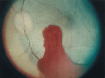
FIGURE 34–1 Vitreous hemorrhage induced by an Nd:YAG (1064 mm) Q-switched laser in a rhesus monkey eye. (Courtesy of the Medical Research Department, Walter Reed Army Institute of Research, San Antonio, Texas.)
• Almost all accidents could have been avoided by following standard laser safety practices; most victims did not wear eye protection because of lack of comfort.
• The injuries typically occurred during alignment of the laser beam or other adjustment procedures.
• Most of the reported injuries have been parafoveal, indicating that the victim was not looking directly into the laser source.
• Occasionally, the beam took an unexpected path, for example, unintentionally reflected by a mirror or nearby object such as photographic paper or a plastic membrane.
• Unexpected discharge of the laser device is rare.
• Almost all of the reported injuries were caused by short-pulse lasers, mostly Nd:YAG, operating in the visible and near-infrared spectrum and emitting a few mJ to tens of mJ per pulse of duration in the tens of nanoseconds range.
The clinical findings include the following.
• The victim experiences a sudden and severe disturbance of vision in one eye, often preceded by a visible flash of bright-colored light; occasionally, there is an audible “pop”.
• The visual acuity is markedly decreased, commonly 20/200 or worse.
• Visual field defects are present.
Stay updated, free articles. Join our Telegram channel

Full access? Get Clinical Tree


