Pharmaceutical Agents


Pharmaceutical Agents


The Prescription/Patient Compliance

Jill C. Autry


THE PRESCRIPTION DOS AND DONT’S: PRESCRIBING GUIDELINES (SEE FIGS. 5-1, 5-2 AND 5-3 AND TABLES 5-1 AND 5-2)


  • Do write legibly.


  • Do check drug spelling; many drug names look alike.


  • Do include dosage form, quantity, and strength of medication.


  • Do specify length of treatment on acute care prescriptions.


  • Do use the most economical size package on chronic care prescriptions.


  • Do make sure the patient understands all special instructions, for example


    • Refrigeration


    • Shake suspensions


    • Proper instillation techniques


  • Do specify the symptoms that require “as needed” treatment, for example


    • Itch


    • Pain


    • Redness


  • Do use only metric measurement. DO NOT use household or apothecary measurement systems.


  • Do not preprint your DEA number on your prescription pad.


  • Do use tamper resistant prescription pads where required by law.


  • Do not leave prescription pads unattended on counter tops.


IMPROVING PATIENT COMPLIANCE

One of the most difficult things that any physician faces in managing the patient with chronic disease, be it systemic hypertension, elevated cholesterol, diabetes, or, as in ophthalmic cases, glaucoma and dry eye, is patient compliance with drug therapy.

Use the following recording and education sheets to assist your patient to utilize their medications more effectively (Figs. 5-4, 5-5 and 5-6).


Ophthalmic Dyes

Jill C. Autry

Ophthalmic dyes are among the most useful diagnostic agents used in the management and detection of disorders of the visual system. The most common uses of these agents and their properties are listed in Tables 5-3, 5-4, 5-5, 5-6, 5-7 and 5-8 and Figure 5-7.


Anesthetic Agents

Jill C. Autry

Clinical procedures that may induce patient discomfort should always include the use of an appropriate anesthetic agent. These drugs can vary in their ability to decrease pain, duration of effect, and potential to induce allergic and toxic reactions. It is important to select the most appropriate agent with adequate efficacy and minimal adverse effects (Tables 5-9, 5-10, 5-11, 5-12 and 5-13). If a patient
reports an allergy to an anesthetic agent in one chemical class, an agent from the alternate class can be considered with caution. For example, if a patient reports an allergy to tetracaine (an ester compound), a compounding pharmacist can prepare topical lidocaine (an amide compound) for ocular use.


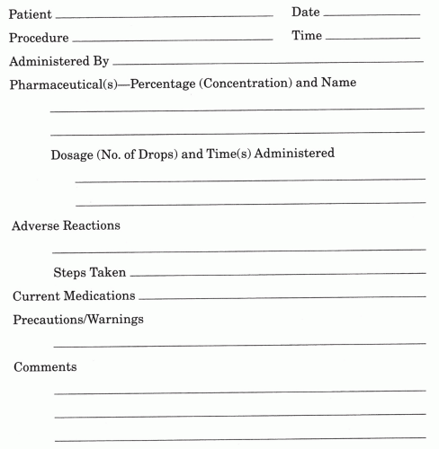
Figure 5-1. Sample drug record—diagnostic.


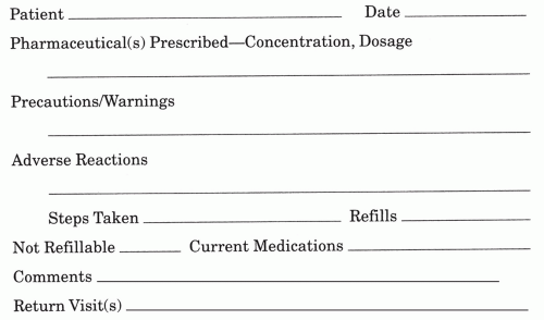
Figure 5-2. Sample drug record—therapeutic.


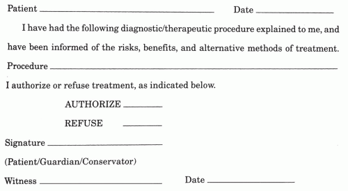
Figure 5-3. Sample consent form.


TABLE 5-1 Lati Abbreviations Commonly Used in Ophthalmic Prescription Writing


Latin


Abbreviation


Meaning


Ad


Ad


Up to, to


Admove


Admov.


Apply


Alternis horis


Alt. hor.


Every other hour


Ante


A


Before


Ante cibos


ac


Before meals


Bis in di’e


bid


Twice a day


Capsula


caps


Capsule


Cum


[C with bar above]


With


Diebus alternis


Dieb. alt.


Every other day


Gram


g, gm


Gram


Gutta


gt, gtt


A drop


Hora


h


An hour


Hora somni


hs


At bedtime


Nocte


noct.


At night


Oculo utro


O.U.


Each eye


Oculus dexter


O.D.


Right eye


Oculus sinister


O.S.


Left eye


Per os


po


By mouth


Pro re nata


prn


When needed


Quaque


q


Each, every


Quaque hora


qh


Every hour


Quater in di’e


qid


Four times a day


Recipe


Rx


Take, you take


Signatura


Sig.


Write, you write


Sine


[s with bar above]


Without


Solutio


Sol.


Solution


Tabella


tab


Tablet


Ter in di’e


tid


Three times a day


Unguentum


ung.


Ointment


Ut dictum


Ut dict.


As directed


Unus


i


One


Duo


ii


Two


Tres


iii


Three


Quattour


iv


Four


Quinque


v


Five


TABLE 5-2 A “Minimum List” of Dangerous Abbreviations, Acronyms, and Symbols Has Been Approved by Joint Commission (Beginning January 1, 2004, the Following Items Must Be Included on Each Accredited Organization’s “Do Not Use” List)


Set


Item


Abbreviation


Potential problem


Preferred term


1.


1.


U (for unit)


Mistaken as zero, four or cc.


Write “unit.”


2.


2.


IU (for international unit)


Mistaken as IV (intravenous) or 10 (ten).


Write “international unit.”


3.


3.


4.


q.d.,


Q.O.D. (Latin abbreviation for once daily and every other day)


Mistaken for each other.


The period after the Q can be mistaken for an “I” and the “O” can be mistaken for “I.”


Write “daily” and “every other day.”


4.


5.


6.


Trailing zero (X.0 mg)


[Note: Prohibited only for medication-related notations];


Lack of leading zero (.X mg)


Decimal point is missed.


Never write a zero by itself after a decimal point (X mg), and always use a zero before a decimal point (0.X mg).


5.


7.


8.


9.


MS


MSO4


MgSO4


Confused for one another.


Can mean morphine sulfate or magnesium sulfate.


Write “morphine sulfate” or “magnesium sulfate.”


Effective April 1, 2004 (if your organization does not already have additional “do not use” items in place), each organization must identify and apply at least another three “do not use” abbreviations, acronyms, or symbols of its own choosing. (Revised 11/3/03.)


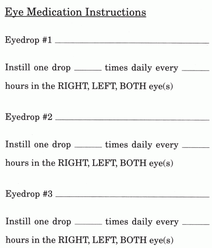
Figure 5-4. Patient medication instruction sheet.


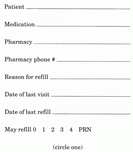
Figure 5-5. Medication refill request form.


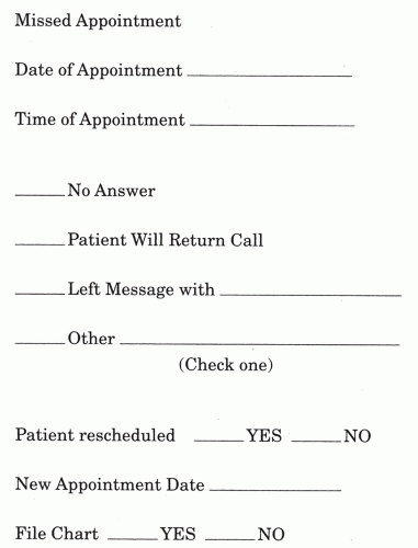
Figure 5-6. Missed appointment record sheet.


TABLE 5-3 Common Uses of Ophthalmic Dyes


FLUORESCEIN


Lacrimal testing (TBUT, Jones test)


Detection of corneal epithelial defects


Detection of penetration of globe (Seidel’s sign)


Applanation tonometry


Contact lens/corneal fitting relationship


Retinal angiography


LISSAMINE GREEN


Dry eye evaluation


FLUOREXON


Evaluation of soft contact lens fit


ROSE BENGAL


Dry eye evaluation


Herpes simplex keratitis


INDOCYANINE GREEN


Angiography


TABLE 5-4 Properties and Characteristics of Fluorescein


CHEMICAL PROPERTIES


C2H12O5Na


Molecular weight = 376.27


Orange-red powder


Yellow in solution


Fluorescence


Yellow-green with blue light in weak alkali solution


Peak absorption: 465-490 nm


Peak emission: 520-530 nm


Fluorescence increases with greater concentrations up to 0.001%


Fluorescence increases with greater pH up to pH 8


At very high concentrations:


Quenching occurs


It dimerizes and polymerizes


Emission shifts to longer wavelengths


CLINICAL CHARACTERISTICS


Stains epithelial defects bright green


Diffuses into intercellular spaces


Will not stain devitalized cells nor mucus


Tear film appears yellow-orange


Can exhibit:


Pseudoflare


Fisher-Schweizer mosaic


Negative staining


Promotes growth of Pseudomonas in solution


Will stain soft contact lenses


TABLE 5-5 Characteristics of Fluorexon


N1 N-bis (carboxymethyl)-aminoethyl-fluorescein tetra-sodium salt


Molecular weight = 710


Stains epithelial defects, devitalized cells, and mucin (less than fluorescein or rose bengal)


Fluorescence


Less brilliant than fluorescein


Does not increase linearly with increasing concentration


Does not stain most soft contact lenses


May stain high water (>60%) contact lenses


Promotes the growth of Pseudomonas


TABLE 5-6 Characteristics of Rose Bengal


4,5,6,7-tetrachloro-2’,4’,5’,7’-tetraiodofluorescein sodium


Stains devitalized cells and mucin a brilliant red


Stains the nucleus more than cytoplasm


Higher concentrations stain more distinctly and highlight more subtle defects


Stings upon instillation


Antiviral properties


Stains a line along the ciliary margin on conjunctival tarsus in normal eyes


TABLE 5-7 Comparative Staining Characteristics of Fluorescein, Rose Bengal, and Fluorexon


Epithelial defects


Devitalized cells


Mucin


Fluorescein


++


0


+


Rose bengal


0


++


++


Fluorexon


+


+


+


Lissamine green


0


++


+


TABLE 5-8 Topical Products


Product


Concentration


How supplied


FLUORESCEIN


Fluorescein (Alcon, Iolab)


2%


1, 2, and 15 mL


Fluorocaine (Akorn)


0.25% fluorescein sodium, 0.1% proparacaine hydrochloride, 0.01% thimerosal preservative


5 mL


Flucaine (Pharmafair, Inc)


0.25% fluorescein sodium, 0.50% proparacaine hydrochloride, 0.01% thimerosal preservative (povidone, boric acid polysorbate 80)


5 mL


Fluress (Sola/Barnes-Hind)


0.25% fluorescein sodium, 0.4% benoxinate hydrochloride, 1% chlorbutanol, povidone buffers


5 mL


Ful-Glo (Sola/Barnes-Hind)


0.6-mg strips


300 strips


Fluor-1-Strip (Wyeth-Ayerst)


9-mg strips (boric acid polysorbate 80, 0.5% chlorbutanol)


300 strips


Fluor-1-Strip AT (Wyeth-Ayerst)


1-mg strips (boric acid polysorbate 80, 0.5% chlorbutanol)


300 strips


Fluorets (Akorn)


1-mg strips


100-1,300 strips


FLUOREXON


Floresoft (Holles)


0.35%


0.5-mL pipettes


LISSAMINE GREEN


1-mg strips


100 strips


ROSE BENGAL


Rose Bengal 1% (Akorn) Americil


1% solution (povidone, sodium borate, PEG P-isoc tylphenolio, 0.01% thimerosal)


5 mL


Rose Bengal (Sola/Barnes-Hind)


1.3-mg strip


100 strips


Rosets (Smith & Nephew)


1.3-mg strip


100 strips


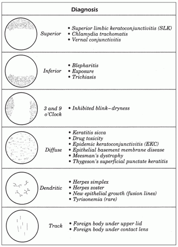
Figure 5-7. Common corneal fluorescein patterns.


TABLE 5-9 Physiochemical and Pharmacologic Properties of the Injectable Local Anesthetics


Physiochemical properties


Pharmacologic properties


Agent (Trade Name)


pKa (25°)


Partition coefficient*


Percent protein binding (%)


Onset (min


Relative potency


Duration (h†)


Concentration (%)


ESTERS Procaine (Novocain)


8.9


0.02


6


Slow (15-25)


1


Short (0.5-1)


1-2


Tetracaine (Pontocaine)


8.5


4.1


76


Slow (20-30)


8


Long (3-5)


0.25


AMIDES


Prilocaine (Citanest)


7.9


0.9


55


Fast (5-15)


2


Moderate (1-3)


1-3


Lidocaine (Xylocaine)


7.9


2.9


64


Fast (5-15)


2


Moderate (1-3)


0.5-2


Mepivacaine (Carbocaine)


7.6


0.8


78


Fast (5-15)


2


Moderate (1-3)


1-2


Bupivacaine (Marcaine)


8.1


27.5


96


Moderate (10-20)


8


Long (3-5)


0.25-0.75


Etidocaine (Duranest)


7.7


141


94


Fast (5-15)


6


Long (3-5)


0.5-1


* Partition coefficient is a measure of lipid solubility.

Onset and duration times seen with epidural injection.


Modified from Covino BG. Pharmacology of local anesthetics. Ration Drug Ther 1987;21:1-9, and Local anesthetics. In: Facts and Comparisons, St. Louis: Lippincott, 1990: 691-694.


TABLE 5-10 Commonly Used Topical Ocular Anesthetics


Drug (Trade Name)


Concentration (%)


Onset (s)


Duration (min)


Maximum dose* (drops


Benoxinate (Fluress)


0.4


20


10


Cocaine§


1.0-10


30


12


20 mg (about 5 drops to each eye of a 4% solution)


Proparacaine


(various)


(Fluoracaine)


(I-Parescein)


0.5


15


14


10 mg (about 20 drops to each of a 0.5% solution)


Tetracaine (various)


0.5


20


10


5 mg (about 10 drops to each eye of a 0.5% solution)


(Pontocaine eye)||


* Suggested maximum total topical doses. (Based on theoretical calculations from Lyle WM, Page C. Possible adverse effects from local anesthetics and the treatment of these reactions. Am J Optom Physiol Opt 1975;52:736.)

Assumes approximately 20 drops/mL of solution.

Combined with 0.25% fluorescein sodium.

§ Must be prepared from a powder.

|| Ointment, 0.5%.


TABLE 5-11 Central Nervous System Toxicities of Local Anesthetics


EARLY EXCITATORY EFFECTS


Restlessness


Anxiety


Dizziness


Tinnitus


Miosis


Tremors


Convulsions


DEPRESSIVE EFFECTS (MAY OR MAY NOT BE PRECEDED BY THE EXCITATORY SYMPTOMS)


Drowsiness


Sedation


Generalized CNS depression


Unconsciousness


Coma


Apnea and respiratory depression


Death from respiratory arrest


TABLE 5-12 Cardiovascular Symptoms of Toxicity from Local Anesthetics


Hypertension and tachycardia (associated with the CNS excitatory phase)


Myocardial depression


Decreased cardiac output


Peripheral vasodilation


Hypotension


Bradycardia


Methemoglobinemia (seen only with prilocaine)


Heart block


Ventricular arrhythmias


Circulatory collapse


TABLE 5-13 Ocular Toxicity from Acute Administration of Topical Ocular Anesthetics


Mild stinging, burning


Vasodilation


Shortening of the tear breakup time


Decreased reflex tearing


Decreased blinking


Corneal edema


Decreased epithelial mitosis and migration


Slowed epithelial healing


Punctate epithelial keratitis


Epithelial desquamation


Allergic reactions of the lids and conjunctiva


Dilating Agents and Procedures

Jill C. Autry

Of all the tests that we, as primary eyecare providers, perform to assess the ocular health of our patients, there is probably no more important test than the dilated fundus examination.

This section provides a quick reference to the procedures and medications that will improve the efficiency of this all-important procedure (Tables 5-14 and 5-15).

Prior to dilation, the clinician must assess the potential for angle closure. This can be done by gonioscopy or by the slit-lamp estimation procedure as seen in Table 5-16. Of course, patients most likely to develop angle closure are older, phakic patients with emmetropic to hyperopic prescriptions. See also Tables 5-17 and 5-18.


TABLE 5-14 Indications for Dilation


1.


Any new patient


2.


Established patient every 2-4 y


3.


Any patient with flashes or floaters


4.


Unexplained vision loss


5.


Any patient with progressive retinal disease (e.g., macular degeneration, diabetic retinopathy)


6.


Any patient with systemic disorders that can affect the eye (e.g., systemic hypertension)


7.


History of ocular trauma


8.


History of chronic uveitis


9.


Any child under 6 y (with cycloplegia)


10.


Once yearly if history of ocular surgery


11.


Moderate to high myopia:


a. 3-7 D of myopia—at least every 2 y


b. >8 D of myopia—at least yearly


12.


Any patient with history of peripheral retinal degeneration


13.


Any patient with developing cataract


14.


All hyperopes or anisometropes (include cycloplegic refraction)


TABLE 5-15 Mydriatic Pharmaceuticals for Dilation


Drug


Maximal dilation


Duration


PARASYMPATHOLYTIC AGENTS


Tropicamide


0.5%


20-40 min


4-8 h


1.0%


6-8 h


Cyclopentolate


0.5%


30-60 min


12-24 h


1.0%


12-36 h


Homatropine


2.0%


40-60 min


24-72 h


5.0%


36-72 h


Scopolamine


0.25%


20-30 min


3-7 d


Atropine


1.0%


30-40 min


7-10 d


SYMPATHOMIMETIC AGENTS


Phenylephrine


2.5%


20-30 min


4-6 h


10%


Hydroxyamphetamine


1%


25-35 min


4-6 h


TABLE 5-16 Van Herick Filtration Estimation Guide


A/C depth at limbus compared to corneal thickness at limbus


Estimated grade of angle


1/2-1


Grade 4


1/4-1/2


Grade 3


=1/4


Grade 2


<1/4


Grade 1


CLOSED


Grade 0


TABLE 5-17 Examination Procedures in Relation to the DFE


I.


Perform these procedures prior to dilation:


1. Case history


2. Best corrected acuity


4. Near-point accommodation and stereopsis


5. External evaluation and pupil testing


6. Biomicroscopy


7. Tonometry


8. Gonioscopy if indicated


II.


Perform these procedures during dilation:


1. Color vision


2. Visual field


3. Frame selection


4. Patient education


III.


Perform these procedures after dilation:


1. Repeat retinoscopy and subjective to R/O latent hyperopia


2. Binocular indirect ophthalmoscopy


3. Slit lamp of lens and vitreous chamber with 60, 78, or 90 D lens


TABLE 5-18 Example of the “Round-Robin” Approach to Patient Scheduling for Dilation


Morning schedule*


Procedure


8:30-8:55 AM


Predilation examination of new patient A


8:55-9:20 AM


Predilation examination of new patient B


9:20-9:30 AM


Dilated exam and dismissal of patient A


9:30-9:40 AM


Brief follow-up examination of patient C (e.g., contact or glaucoma IOP recheck)


9:40-9:50 AM


Dilated exam and dismissal of patient B


9:50-10:15 AM


Predilation examination of new patient D


10:15-10:40 AM


Predilation examination of new patient E


10:40-10:50 AM


Dilated exam and dismissal of patient D


10:50-11:00 AM


Follow-up examination of patient F


11:00-11:10 AM


Dilated exam and dismissal of patient E


11:10-11:35 AM


Predilation examination of new patient G


11:35-11:50 AM


Follow-up examinations as needed and return morning phone calls


11:50-noon


Dilated exam and dismissal of patient G


* Afternoon schedule may repeat morning or use for specialty appointments. Examination times may vary from doctor to doctor. Delegation of tests and an adequate number of examination rooms is necessary to follow this type of scheduling.


Diagnostic Agents and Procedures for the Management of Accommodative Disorders

Jill C. Autry

The use of diagnostic pharmaceuticals during the examination procedure is necessary in a number of situations. This section provides information regarding the appropriate use of pharmaceutical agents for the diagnosis of accommodative disorders (Tables 5-19, 5-20, 5-21, 5-22, 5-23 and 5-24 and Fig. 5-8) and accommodative esotropia (Table 5-25 and Fig. 5-9).


TABLE 5-19 Indications for Cycloplegic Refraction


1. Strabismus (particularly esotropia)


2. Amblyopia


3. Anisometropia


4. Pseudomyopia


5. Hyperopia associated with esophoria or a lag of accommodation


6. Unstable end point on static retinoscopy


7. For the uncooperative child during static retinoscopy


8. Preparation for refractive surgery


Ophthalmic Lubricants

Jill C. Autry

Ophthalmic lubricants represent the cornerstone of the management of ocular surface disease. New formulations of these all-important products have recognized the importance of limiting preservative toxicity while enhancing epithelial cell growth. Tables 5-26, 5-27, 5-28, 5-29 and 5-30 list the most common products.


TABLE 5-20 Comparison of Cycloplegic Agents


Drug


Dosage


Onset of cycloplegia


Duration of cycloplegia


Tropicamide 1%


1 drop, repeat after 5 min


20-30 min


4-8 h


Cyclopentolate 0.5% 1.0%, 2%


1 drop, repeat after 5 min


20-45 min


8-24 h


Homatropine 5%


1 drop, repeat after 5 min


30-60 min


24-48 h


Scopolamine 0.25%


1 drop, repeat after 20 min


30-60 min


5-7 d


Atropine


0.5% ointment


1/4”ointment at bedtime for 3 d prior to exam


30-60 min


10-14 d


1.0% solution


1 drop tid × 1 d prior to exam


TABLE 5-21 Efficiency of Cycloplegics


Drug


% Efficiency


1% Atropine


100


1% Cyclopentolate


92


1% Tropicamide


80


5% Homatropine


54


TABLE 5-22 Ocular Side Effects of Cycloplegic Agents


Allergic contact dermatitis


Angle-closure glaucoma


Elevation of IOP with open angles


TABLE 5-23 Dose-related Systemic Side Effects of Atropine


Dose


Effects


0.5-2 mg (1-4 drops 1% solution)


Tachycardia


Dry mouth


Mydriasis/cycloplegia


5 mg (10 drops 1% solution)


The above plus:


Speech disturbance


Restlessness


Confusion


Hot, dry skin


Decreased GI motility


Urinary retention


>10 mg (over 20 drops 1% solution)


The above plus:


Hyperexcitability


Hallucination


Coma


Convulsion


Death


TABLE 5-24 Side Effects Associated with Topical Cholinesterase Inhibitors


OCULAR


Ciliary body


Accommodative spasm*


Anterior movement of the lens-iris diaphragm


Breakdown of the blood-aqueous barrier


Decreased anterior chamber depth


Conjunctiva


Drug-induced cicatrizing conjunctivitis


Hyperemia


Corneal toxicity


Increased intraocular pressure (paradoxical)


Lens


Cataract formation (particularly anterior subcapsular cataracts)


Lids


Allergic blepharoconjunctivitis


Depigmentation of the skin (reversible)


Twitching of orbicularis oculi


Pupil


Iris cysts*


Miosis


Retina


Increased peripheral vitreoretinal traction


SYSTEMIC


Cardiac


Arrhythmia


Bradycardia


Gastrointestinal*


Abdominal cramps


Diarrhea


Nausea


Headache


Pulmonary


Bronchospasm


Upper respiratory congestion


Lacrimation


Reduced plasma cholinesterase levels


Reduced breakdown of succinylcholine, procaine, and tetracaine


Rhinorrhea


Urinary incontinence


* More common in children.

More common in elderly.


TABLE 5-25 Characteristics of Accommodative Esotropia


1. Type of onset: Gradual and intermittent


2. Time of onset: Typically 2.5-4 y of age with a range from 4 mo to 7 y


3. Typically large angle of 25-40 degrees


4. Concomitant


5. Normal correspondence


6. Seldom have significant amblyopia


7. Refractive type:


Normal AC/A


Hyperopia between 2.00 and 7.00 D, with mean of 4.50 D


8. Nonrefractive type:


High AC/A ratio (>6:1)


Hyperopia between 0.50 and 4.50 D, with mean of 2.00 D


9. Can have a combination of refractive and nonrefractive components


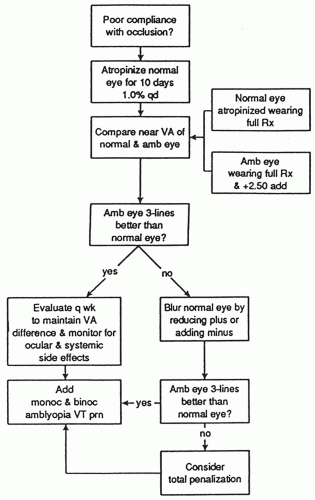
Figure 5-8. Management of amblyopia with penalization.

Only gold members can continue reading. Log In or Register to continue

Stay updated, free articles. Join our Telegram channel

Jul 21, 2016 | Posted by in OPHTHALMOLOGY | Comments Off on Pharmaceutical Agents

Full access? Get Clinical Tree

Get Clinical Tree app for offline access