CHAPTER 19 Phaco complications
Introduction
Operative complications can occur at any stage of cataract surgery, anywhere from the administration of local anesthesia right up to the injection of intracameral antibiotics. There are many bear traps and pitfalls along the way that can trip up the inexperienced, unwary, or inattentive surgeon. We need to better understand precisely what those pitfalls are and to anticipate and react early to them. If we do not, then nothing changes. We carry on getting the same complications with the same incidence for the same reasons, ad infinitum. Without wanting to sound too sanctimonious I believe that we need to approach every complication as an opportunity to learn something new. Each event then adds cumulatively to our library of cognitive and technical skills.
This chapter is intended to provide a clear guide to the practical management of the more common and important intraoperative complications and will cover them in sequence through the different stages of phaco surgery.
Local anesthesia
Peribulbar hemorrhage
This can occur with any type of infiltrative anesthesia and is usually fairly immediate but can occasionally be delayed until later during surgery. The orbit becomes tense and incompressible but will usually soften fairly quickly over 10–20 minutes if it is due to the far more common venous bleed. The lids can become tight and difficult to open but again will usually settle down given a little time and some direct orbital pressure. An oculopressive balloon can help limit bleeding and lower the orbital tension. Occasionally the orbit will remain tense, in which case surgery should be abandoned because the risk of chamber collapse, iris prolapse, posterior capsule rupture, and vitreous loss is high. Most patients are relieved that you are putting safety first, once they have understood the risks of continuing. In practice only a small minority (around 5%) of patients with peri- or retrobulbar hemorrhage will need to have their surgery rescheduled1.
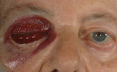
Very rarely an orbital arterial bleed may occur. This has happened to the author only once in 20 years of eye surgery. In this case there is rapid and dramatic proptosis accompanied by a rock hard orbit and infiltrative staining of skin and conjunctiva by pressurized blood (Fig. 19.1). Despite intravenous acetazolamide and mannitol plus lateral canthotomy/cantholysis the visual prognosis following arterial orbital hemorrhage is often poor.
Needle injury
Any needle can damage an extraocular muscle, perforate the globe or, if it is long enough, enter the optic nerve via the intraconal space. High risk orbital apex injections have now largely been abandoned and superceded by safer topical, peribulbar, and subTenon’s anesthesia. Consequently subarachnoid infiltration and brainstem anesthesia are almost unheard of today. The incidence of single penetration or double perforation with peribulbar anesthesia is less than 1 in 20002 but increases to around 1 in 150 with larger myopic eyes3. If a short disposable sharp 25 mm needle is used and the surgeon is familiar with the anatomy of the orbit then it is difficult to inadvertently enter a normally sized eye without adequate warning signs. First, you will meet with a significant increase in resistance to the passage of the needle as you tangentially catch the sclera. This is often associated with some pain. However the key sign is that the impaled eye rotates with advancement of the needle. If any of these three things happen then stop, withdraw, and enter more peripherally with the needle directed parallel with the orbital wall.
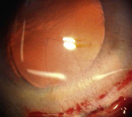
Incisions
Problems with wound construction and architecture are fairly common as a reproducibly ‘perfect’ incision is difficult to achieve. The ‘Rule of Too’s’ describes most of the root causes: too short, too long, too wide, too narrow, too central, too peripheral, too superficial, or too deep.
If it is too short then intraoperative wound leakage with chamber instability will be a problem, as well as an increased risk of iris prolapse and a leaking wound at the end that requires suturing. It will be clear from the start that there is a problem and the best option is to suture the wound and make a better incision at a different site.
A peripheral wound will have breached the conjunctival insertion and is therefore prone to progressive conjunctival ballooning. This leads to fluid pooling on the cornea with resulting poor visibility from distortion and reflections. This is best resolved by extending the conjunctival incision and retracting the back edge of the conjunctiva so that the incisional outflow is no longer sequestered into the subconjunctival space. Firmly massaging already ballooned conjunctiva with a squint hook effectively disperses excess fluid.
Wounds that start peripherally also usually enter the chamber peripherally, so they are additionally prone to iris prolapse. Best to reposit the iris, suture the wound, and make a new incision.
A wound that is too long or too central makes the rhexis difficult and usually undersized as well as eccentric. It also leads to corneal distortion when pointing the phaco tip downwards (so-called ‘roofing’) causing impaired visibility. Such distortion can even tear the back edge of the roof of the incision. A long wound as well as a narrow one will increase the likelihood of a corneal burn, despite modern power modulations such as pulse, burst, and torsional modes. Most significant wound burns result in tissue contraction and wound-gape and need a mattress suture to close them on the table, which may be difficult. The majority will settle over subsequent weeks with conservative management on topical steroids and antibiotic cover. Very rarely scleral patching may be required.
A small dehiscence of Descemet’s membrane related to the internal front edge of the main wound is fairly common. This produces a hinged flap which spontaneously falls back into place and is of no consequence. A larger flap can, on most occasions, be persuaded to lie flat again. If not then an air bubble, SF6 gas, viscoelastic, or rarely suturing have all been used to successfully re-attach hanging flaps. The problem arises when a sizeable flap is stripped off and then completely detaches, never to be seen again. This is rare. The defect is usually triangular in shape with its base the width of the incision and its apex towards the center of the cornea (Fig. 19.2). Nothing to do here except wait and see how well the remaining endothelial cells respond to the greater demand on them. If over one fifth of the endothelium is gone then grafting is likely to be needed. Wait at least 3 months before considering endothelial grafting since slow recovery is the usual course with smaller defects.
When using scleral tunnels caution is needed to avoid cutting the groove too deep otherwise the choroid and suprachoroidal space can be exposed. The sclera should be sutured and a new site used. Scleral tunnels also carry a risk of tunnel hemorrhage, which occurs in up to 10% of such incisions. These are usually obvious at the time of surgery but can be delayed and associated with large postoperative hyphemas. Hemorrhage can be limited by rapid tamponade using viscoelastic. Fastidious hemostasis at the time of surgery is mostly successful in avoiding further problems.
Capsulorrhexis
The correct sizing of the capsulorrhexis is important not only for trapping the anterior edge of the implant optic with an overlapping rim of capsule, but also for avoiding a number of problems and complications. A rhexis that is too small increases the risk of capsular block and possible posterior capsule rupture during hydrodissection. It also is more likely to suffer a secondary radial tear from the phaco tip or second instrument. It should be enlarged at the time and not left to chance. An oversized rhexis is also problematical. The nucleus is likely to tumble out during hydrodissection and also the optic of the IOL will be prone to forward prolapse or decentration from asymmetric capture by the large rhexis. The usual cause of an oversized rhexis is an oversized pupil. We seem to be irresistibly drawn to the edge of the pupil. Nothing you can do about an oversized rhexis except avoid it in the first place.

Stay updated, free articles. Join our Telegram channel

Full access? Get Clinical Tree

