Uno et al. [14] used rats and examined the metabolic activities in proximal cell bodies that received the signal caused by peripheral nerve injury. They focused on the changes in the production of growth factor associated protein-43 (GAP-43), which is thought to play an important role in regeneration. The study showed that GAP-43 mRNA expression increased in the cell bodies of the nucleus ambiguus motor neurons after recurrent laryngeal nerve transection. The expression peaked at 1 week after transection and began to decrease at approximately 2 weeks after transection. This peak period of GAP-43 mRNA expression was different from that in the nuclei of other peripheral nerves after transection. The regenerative ability of the laryngeal nerve was much lower than that of other nerves.
6.3 Artificial Nerve Conduit
In the regenerative process of injured peripheral nerves, an environment favorable to regeneration is important at as early a stage as possible to achieve good results. Thus, we created an artificial nerve conduit, a polyglycolic acid (PGA) tube, based on a basic concept of in situ tissue engineering [15]. The surface of this tube is covered with a mixture of porcine collagen type I (70 %) and collagen type III (30 %). A collagen had low antigenicity due to removal of telopeptide. The tube was then filled with the same collagen and treated dehydrothermally at 140°C at a pressure of 10 Pa for 24 h in order to cross-link the collagen molecules. PGA is a high-molecular-weight compound with the simplest aliphatic polyester polymer. It is hydrolyzed in a living body and completely degrades in approximately 4 months. On the other hand, it maintains its shape for a certain period of time.
We have prepared several types of PGA tubes to date and conducted experiments on them. With a goal of clinical application, we created the current type that can be mass produced and has superior physical strength (Fig. 6.2).
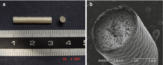
Fig. 6.2
Artificial nerve conduit. (a) Polyglycolic acid (PGA) tube in longitudinal and cross-sectional views. (b) Image of PGA tube by scanning electrochemical microscope. The outer and inner surfaces are coated with amorphous collagen layers and filling its interior with a collagen sponge
Studies have been conducted on rats, cats, and dogs to regenerate peripheral nerves after experimental resection. PGA tubes filled with collagen were used in these studies. The results in dogs indicated that the recurrent laryngeal nerve can have functional regeneration after resection [9, 11] (Figs. 6.3, 6.4, and 6.5).
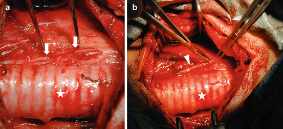
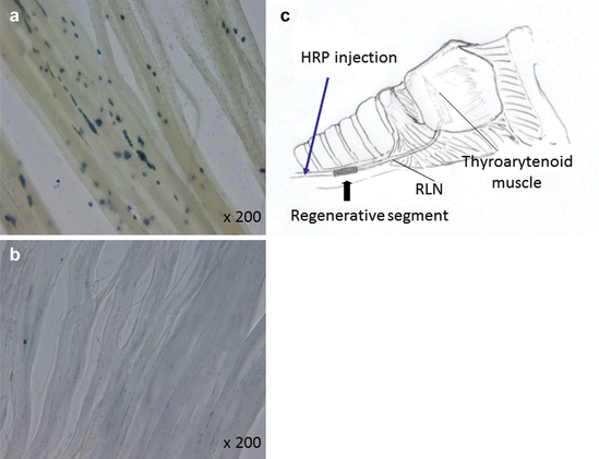
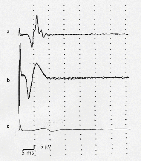
Fig. 6.3
Intraoperative views of canine model. (a) After resection of RLN, (b) Reconstruction of nerve defect by PGA tube. White arrow: Recurrent laryngeal nerve (RLN) ends (10 mm defect); white arrow head: Reconstruction of RLN by PGA tube; white asterisk: Trachea
Fig. 6.4
Recovery of the axonal transport of severed recurrent laryngeal nerve (RLN). (c) At 6 months after operation, wheat germ agglutinin-horseradish peroxidase (HRP) was injected on the proximal side of the reconstructed RLN. Nerve terminals in cricoarytenoid muscles were stained with HRP on PGA tube side (a) but were not stained on auto-nerve implantation side (b)
Fig. 6.5
Electrophysiological assessment: Compound muscle action potentials (CMAPs) from cricoarytenoid muscles, 6 months after operation. (a) PGA tube reconstructed side, (b) nonoperated side (normal control), (c) auto-nerve graft side
6.4 Clinical Application of PGA Tube for Nerve Regeneration
Based on the aforementioned research results, we began to use the PGA tube in clinical application for nerve regeneration in 2002 after the review and approval by the ethics committees of the Department of Otolaryngology, Head and Neck Surgery, at Kyoto University Hospital in Kyoto and the Department of Otolaryngology, Head and Neck Surgery, at Kitano Hospital in Osaka, Japan.
6.4.1 Recurrent Laryngeal Nerve
As mentioned earlier, the recurrent laryngeal nerve is a nerve whose functional regeneration is most difficult to achieve. We have used PGA tubes in six patients to date, of whom only one showed movement of the vocal cords. This patient was a 54-year-old man who had laryngeal cancer with primary glottic cancer. He had undergone radiation therapy and laser surgery and achieved local control. However, he had cervical metastasis with invasion of the recurrent laryngeal nerve. Therefore, 1 cm of the right recurrent laryngeal nerve was resected and a PGA tube was used for nerve regeneration. Three months after regeneration, the patient had vocal cord movements and functional regeneration of the recurrent laryngeal nerve.
Stay updated, free articles. Join our Telegram channel

Full access? Get Clinical Tree


