Pediatric Traction Retinal Detachments
 Diverse disease processes may create traction retinal detachments in the pediatric age group. Special approaches are required to manage these difficult problems. These young patients have many years ahead of them and require the most aggressive attempts at restoration of vision. Patients in the pediatric age group having retinal detachments of several years’ duration can have remarkable visual recovery, and this emphasizes the need to proceed with such cases. By contrast, the very young patient, especially with retinopathy of prematurity, is a high medical risk patient requiring careful assessment of the risk-benefit ratio of surgery. From the ocular standpoint, unilateral disease in the pre-6-year-old patient has an extremely high incidence of amblyopia, diminishing the visual impact of successful surgery.
PERSISTENT HYPERPLASTIC PRIMARY VITREOUS, PERSISTENT FETAL VASCULATURE
Persistent hyperplastic primary vitreous is also known as persistent fetal vasculature (PFV). It is usually a unilateral phenomenon accompanied by a smaller eye (1,2). The almost uniform incidence of amblyopia means that these cases should be operated on early (3). An additional argument for early surgery is the prevention of long-term traction detachment and pupillary block chamber (4,5). This condition is usually recognized early in life, and the patient should be operated upon if a traction detachment is recognized, the cataract is sufficient to cause visual loss and amblyopia, or there is shallowing of the anterior chamber secondary to pupillary block. The embryological explanation for the syndrome is the lack of regression of the primary vitreous and hyaloid vasculature, although the primary cause is still unclear. Bilateral cases in males are usually associated with Norrie’s syndrome. Norrie’s cases should not be operated on because the retina is dysplastic and the vitreoretinal interface cannot be delineated at surgery.
Surgical Sequence and Techniques
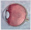
Microincisional vitrectomy with 25-gauge or possible 23-gauge technology is ideal for PFV. The vitrectomy instrument is introduced into the lens substance (Fig. 27.1


Stay updated, free articles. Join our Telegram channel
Full access? Get Clinical Tree
