25-Gauge Vitrectomy
 Sutureless, transconjunctival 25-gauge vitrectomy was introduced by DeJuan and colleagues (1,2) and has rapidly become an accepted tool in the vitreoretinal surgery armamentarium. Sutureless vitrectomy is dependent on two concepts: (a) smaller diameter instruments and therefore smaller sclerotomies and (b) conjunctival displacement before making the transconjunctival sclerotomies so that the conjunctiva covers the sclerotomy after surgery is completed. Trocars are used to place flexible, thin-wall cannulas, resulting in 23.5-gauge sclerotomies. The cannulas are used to maintain alignment between the offset conjunctival incision and the sclerotomy and facilitate finding the small incisions when inserting tools.
CASE SELECTION
Initially it was thought that 25-gauge, sutureless vitrectomy was only indicated for epimacular membranes, macular holes, vitreomacular traction, and surgery for retinal venous occlusion. Early on, many surgeons thought 25-gauge surgery was inappropriate for vitreous hemorrhages, rhegmatogenous retinal detachments, proliferative vitreoretinopathy (PVR), diabetic traction retinal detachments, or giant breaks. Experience has shown, however, that 25-gauge surgery is ideal for vitreous hemorrhages, rhegmatogenous retinal detachments, PVR, and giant breaks and is applicable for diabetic traction retinal detachments as well. Few surgeons now believe that branch vein decompression (sheathotomy) and radial optic neurotomy (RON) are effective procedures, making 25-gauge surgery for these procedures a moot point except in the context of vitrectomy alone for the reduction of macular edema via vascular endothelial growth factor (VEGF) reduction in the macula and increased oxygen tension in the vitreous cavity.
Dislocated lens material (dropped nucleus) and removal of intraocular foreign bodies (IOFBs) require one 20-gauge sclerotomy because dense membranes have too much flow resistance for small-lumen instruments and IOFBs are too large. The technique of combining two 25-gauge sutureless incisions with one 20-gauge sutured incision has been described as 20/25 vitrectomy by the authors.
Patients with prior or anticipated glaucoma- filtering procedures are ideal candidates for 25-gauge surgery. Patients with severe dry eyes, ocular surface disorders, and scarred conjunctiva are excellent candidates for 25-gauge surgery as well.
TROCAR-CANNULA SYSTEM
The purpose of the trocar is to make a 25-gauge sclerotomy and enable simultaneous insertion of flexible 23.5-gauge selfretaining cannula that fits over the trocar (Fig. 9.1). The conjunctival incision is intentionally displaced from the scleral incision so that the two incisions will not be aligned after cannula withdrawal and the conjunctiva will cover the sclerotomy (Fig. 9.2). Repeated insertion and withdrawal of tools is accomplished through the cannula, which maintains the alignment of the conjunctival and scleral incisions and protects the wounds. The conjunctiva should be displaced using a cotton-tip applicator by gently moving it anteriorly or circumferentially or some combination thereof (Fig. 9.3). An attempt should be made to avoid conjunctival and scleral vessels to reduce postoperative subconjunctival hemorrhages. The Alcon noncoring trocar is based on a modified microvitreoretinal (MVR) blade and requires much less insertion force than competitive hypodermic needle-based, coring-type designs. A prospective, consecutive clinical series by the author shows that the Alcon system using fluid-air exchange demonstrates significantly less hypotony than published results with the coring-type trocar. The Alcon trocar cannula usually requires no rotation when inserted. If some resistance is encountered, small-amplitude back and forth rotation will facilitate insertion. The cotton-tip applicator should be handed back to the scrub technician or dropped so that the forefinger of this hand can be used to guide the trocar cannula during insertion. The trocar should be aimed toward a virtual point about 2 mm anterior to the center of the eye or about 2 mm posterior to the lens. The incisions should be made 4 mm posterior to the limbus (Fig. 9.4) unless there is a pars plana abnormality from retinopathy of prematurity (ROP), trauma, or pars planitis; a large choroidal detachment; suprachoroidal hemorrhage; or
high retinal detachment that would necessitate making the incisions through the pars plicata. The 25-gauge incisions may be made 3 mm posterior to the limbus in aphakic eyes. The inferotemporal incision should be made just below the 3 or 9 o’clock position to reduce bleeding and pain but as far as possible from the lower lid so that it will not be displaced if the eye is rotated inferiorly. The superonasal incision is usually made on a virtual line from the lowest point of the bridge of the nose extending through the center of the pupil and then plugged with a special plug made for 25-gauge cannulas. This location reduces tool flexion issues and facilitates peripheral and anterior access. The purpose of the plug is to prevent vitreous prolapse or fluid loss through the port while making the third incision. The superotemporal incision is usually made on a virtual line extending from the lowest point of the supraorbital rim through the center of the pupil again to reduce tool flexion and facilitate anterior and peripheral access.
high retinal detachment that would necessitate making the incisions through the pars plicata. The 25-gauge incisions may be made 3 mm posterior to the limbus in aphakic eyes. The inferotemporal incision should be made just below the 3 or 9 o’clock position to reduce bleeding and pain but as far as possible from the lower lid so that it will not be displaced if the eye is rotated inferiorly. The superonasal incision is usually made on a virtual line from the lowest point of the bridge of the nose extending through the center of the pupil and then plugged with a special plug made for 25-gauge cannulas. This location reduces tool flexion issues and facilitates peripheral and anterior access. The purpose of the plug is to prevent vitreous prolapse or fluid loss through the port while making the third incision. The superotemporal incision is usually made on a virtual line extending from the lowest point of the supraorbital rim through the center of the pupil again to reduce tool flexion and facilitate anterior and peripheral access.
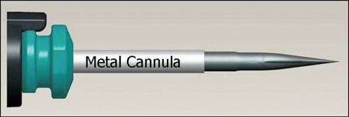 Figure 9.1 ▪ A 25-gauge trocar cannula from Alcon. |
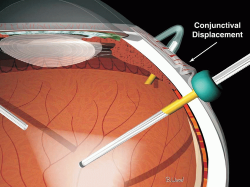 Figure 9.2 ▪ Wound leaks are prevented by conjunctival displacement with respect to the 0.5-mm scleral incision. |
FLUIDICS
Ohm’s law for fluids is directly analogous to Ohm’s law for electricity and teaches that pressure (gradient) is equal to the resistance times the flow. Resistance to flow is proportional to the fourth power of the inner diameter of the lumen. The much higher resistance of 25-gauge cutters and infusion cannulas was initially thought by many to be a major disadvantage. It turns out that increasing infusion pressure to 50 to 60 mm Hg while flow is occurring (dynamic state) and lowering it to 35 to 45 mm Hg (static state) when using forceps, pics, scissors, or the endophotocoagulator (static state) solves the problem. The Alcon Constellation pressurized infusion system and the Accurus Vented Gas Forced Infusion (VGFI) allow rapid and accurate switching between static and dynamic infusion pressures, and the Constellation pressure compensation system directly addresses this problem.
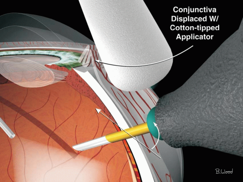 Figure 9.3 ▪ Circumferential or anterior conjunctival displacement with a cotton-tip actuator is done before entry of the trocar cannula. |
The 23/25/27-gauge cutters have more resistance than 20-gauge cutters because of the smaller bore coaxial inner needle as well as the cutter intermittently closing the port during the open-close cycle, thus requiring greater compensation with respect to the vacuum settings used with 20-gauge vitrectomy. The authors use 650 mm Hg maximum setting and proportional (linear) suction and the Constellation system. Fast cutting with 20-gauge cutters is also advantageous because of port-based flow limiting, which results in decreased pulsatile vitreoretinal traction. The smaller bore of the 23/25/27-gauge cutters also produces port-based flow limiting (Fig. 9.5


Stay updated, free articles. Join our Telegram channel
Full access? Get Clinical Tree
