Osteoplastic Frontal Sinusotomy
Kevin C. Welch
INTRODUCTION
The osteoplastic approach to the frontal sinus remains an important alternative in the treatment of chronic inflammatory or neoplastic disease of the frontal sinus. It is interesting to note that what was once the gold standard for treating chronic frontal sinusitis is now considered a surgical technique that many surgeons in training have seldom, if ever, encountered. It is perhaps the lack of familiarity coupled with the success of endoscopic approaches that have caused the osteoplastic frontal sinusotomy to have fallen out of favor as a primary means by which chronic frontal sinusitis is managed. There are several situations, however, in which the approach is appropriate instead of endoscopic approaches. These indications are most aptly characterized in the history and radiologic evaluation of the patient and are described in this chapter.
HISTORY
The patient with chronic frontal sinusitis presents a treatment dilemma to the surgeon since many forms of chronic frontal sinusitis may be appropriately treated by performing an adequate anterior ethmoidectomy and infundibulotomy. Patients falling within this category typically tend not to have widespread mucosal inflammation and polypoid disease or have areas of anatomic obstruction that predispose them to chronic frontal sinusitis. In fact, many would advocate not operating on the frontal sinus to any degree until more conservative approaches fail to resolve symptoms (and disease) referable to the frontal sinus. For the majority that remain, an endoscopic frontal recess dissection will often provide a successful outcome.
Patients who present with a history of sinus surgery, especially previous external sinus surgery (e.g., Lynch frontoethmoidectomy procedures or frontal trephination procedures), should raise suspicion of recalcitrant inflammatory disease or significant postsurgical scarring that may make endoscopic treatment difficult if not impossible. A history of pain in the forehead, edema, erythema, numbness, or change in appearance of the forehead may be indications of significant chronic frontal sinusitis. Lastly, a history of head trauma should be sought and notation made of any surgical repair of facial fractures, as these fractures may distort the natural anatomy of the frontal sinus and most importantly that of the frontal recess.
PHYSICAL EXAMINATION
A complete examination of the head and neck is always advised with a detailed focus on the forehead and periorbital region. The scalp should be inspected for signs of previous surgery or trauma, including evidence of a laceration, bifrontal/coronal incisions, brow incisions, gull-wing incisions, or Lynch incisions. The characteristics
of the overlying skin should be examined for edema, erythema, ballotability, tenderness, or hypesthesia. The examiner should make note of any orbital proptosis, lid lag, anisocoria, or restriction of gaze.
of the overlying skin should be examined for edema, erythema, ballotability, tenderness, or hypesthesia. The examiner should make note of any orbital proptosis, lid lag, anisocoria, or restriction of gaze.
A thorough intranasal endoscopic examination should be performed as well. Endoscopic examination should focus on the extent of infection, edema of the sinonasal mucosa, the presence of polyps and the presence and extent of any previous surgical scarring. The surgeon should look for signs of frontal sinus obstruction, such as a lateralized middle turbinate, retained uncinate process, an undissected agger nasi cell and tumor. If the frontal recess has been previously dissected, examination should be performed with a 70-degree or 45-degree telescope with the intention of exploring the frontal recess for signs of disease.
INDICATIONS
An osteoplastic frontal sinusotomy is indicated for any form of frontal sinus disease that is contained within the boundaries of the frontal sinus, for example, fibro-osseous lesions (fibrous dysplasia, ossifying fibroma), benign neoplasms (e.g., osteoma), defects in the posterior table of the frontal sinus resulting in cerebrospinal fluid (CSF) rhinorrhea, frontal sinus fractures with involvement of the frontal recess, mucoceles, and obstructing frontal recess cells. Previous episodes of Potts puffy tumor may result in frontal sinusitis or scarring that is inaccessible with endoscopic techniques. A review of the radiographic studies may reveal a significant amount of osteitis or osteomyelitis that needs to be drilled down to eliminate a recurrent source of infection. An osteoplastic frontal sinusotomy may also be used in combination with an endoscopic approach to treat any of the aforementioned diagnoses as well if an “above and below” approach is necessary.
CONTRAINDICATIONS
The osteoplastic frontal sinusotomy has relatively few contraindications. An osteoplastic frontal sinusotomy would be contraindicated in cases when a benign or malignant tumor involving the frontal bone extends beyond the limits of the frontal sinus boundaries. In this situation, an osteoplastic approach may not be oncologically sound and consideration should be given for a more definitive procedure (e.g., frontal craniectomy). There are no other absolute contraindications. Other contraindications should be considered as relative. As previously mentioned, this approach may not be appropriate for primary frontal sinus surgery. Additionally, local factors such as previous radiation; significant frontal sinus trauma, especially posterior table trauma; and extent and quality of disease (e.g., inverted papilloma, allergic fungal sinusitis, CSF rhinorrhea) may dictate whether obliteration is performed or if an adjunctive endoscopic approach is necessary. General contraindications related to the patient such as severe cardiopulmonary disease and bleeding diathesis should also be considered relative as well.
Although performing an osteoplastic flap has relatively few absolute contraindications, combining an osteoplastic flap with obliteration can be problematic in certain cases in which the mucosa of the sinus may not be completely exenterated. Frontal sinuses with prominent supraorbital recesses with a narrow anteroposterior diameter can be difficult to instrument in order to remove all the mucosa. Failure to fully remove the mucosa of the sinus increases the likelihood of a postoperative mucocele. Another example is the presence of a supraorbital bony erosion from previous frontal sinus disease. In this instance, the mucosa of the sinus is often adherent to the periorbita, and complete removal of the mucosa is problematic without resecting the underlying periorbita.
PREOPERATIVE PLANNING
The frontal sinus can be approached through multiple incisions, for example, bifrontal/coronal, midforehead, brow, or gull-wing incision. The appropriate approach is dictated by how much exposure is necessary and on certain patient considerations (e.g., age, sex, forehead wrinkles, male pattern baldness, and scars from previous surgery) and the surgeon’s experience or preference. In most cases, the bifrontal/coronal incision provides superior exposure to the entire frontal sinus, minimizes poor cosmetic outcomes, and can be used in men or women. However, this approach is more labor intensive and may be unacceptable for men with male pattern baldness. Other incisions provide adequate exposure but may create unsightly facial scars; therefore, a thorough explanation should be given to the patient before proposing such an approach. Since the bifrontal/coronal approach offers the best exposure, this approach will be described in our surgical technique.
All patients need to undergo imaging as part of the diagnostic evaluation. Computed tomography is considered the best modality for evaluating the bone of the frontal sinus and frontal recess. Evidence of hyperostosis, formation of a mucocele, and areas of dehiscences should be noted and used to determine whether the osteoplastic frontal sinusotomy would be superior to the endoscopic approach. Magnetic resonance imaging is helpful if a tumor is suspected.
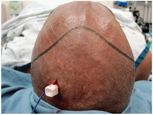 FIGURE 37.1 A proposed incision for the coronal flap. The incision extends from just anterior to the tragus within the preauricular crease to the contralateral side. The incision is drawn anteriorly, and a peak can be designed to help approximation of the scalp flap at the termination of the case. Along the posterior scalp, an incision is made and a skull reference array is attached to the calvarium. |
The method by which the frontal sinus is outlined and entered during the surgical procedure dictates whether a preoperative stereotactic navigation CT scan or a traditional 6-foot Caldwell roentgenogram is necessary. If the surgeon plans to map out the boundaries of the frontal sinus using stereotactic navigation techniques, a 6-foot Caldwell view is unnecessary but may still be obtained as an alternative means for mapping the sinus should the stereotactic navigation system fail to be accurate or for confirmation. If the surgeon chooses to use a more traditional method, two copies of the 6-foot Caldwell view are printed—one for reference and one to be sterilized and used as a template in the surgical field.
SURGICAL TECHNIQUE
The patient is placed in the supine position on the operating table. Once anesthesia is induced, the head of the bed is elevated placing the patient’s head at a level more conducive to performing the initial steps of the operation. Monofilament tarsorrhaphy sutures or corneal shields are placed in order to protect the eyes. The face and scalp are prepared in a sterile fashion.
The bifrontal/coronal incision is drawn through the preauricular crease across the scalp to the contralateral side. The incision is directed more anteriorly such that the incision is made 2 cm posterior to the hairline. A peak in the incision at the vertex of the scalp facilitates realignment at the end of the procedure (Fig. 37.1). The hair may be shaved along the course of this incision. Next, the incision is infiltrated with 1% lidocaine with 1:100,000 epinephrine. Starting in the midline, the skin is incised with the edge of the blade beveled at an angle to the hair follicles. The subcutaneous tissues may be divided sharply or more sparingly with electrocautery, since the hair follicles may be irreparably damaged using the latter technique. Scalp clips are recommended to help with hemostasis (Fig. 37.2)


Stay updated, free articles. Join our Telegram channel
Full access? Get Clinical Tree
