Osteoplastic Frontal Sinus Osteoplasty Combined With Draf 3
Peter John Wormald
INTRODUCTION
The endoscopic modified Lothrop/Draf 3/frontal drillout procedure is currently the most frequently used procedure for extended access to the frontal sinus. Where wide access is required, this procedure has replaced the osteoplastic flap (OPF) as the most common procedure for chronic frontal sinusitis and most benign tumors of the frontal sinus. However, there are still indications for the latter procedure, the most common being large osteomas that fill the frontal sinus (Fig. 36.1), tumors of the frontal sinus with intracranial extension, localized lateral osteitis, laterally based cerebrospinal fluid (CSF) leaks, mucoceles that may be difficult to access through the Draf 3 procedure, and fractures of the frontal sinus. Lesions that extend past the midline of the orbit can be difficult to access especially in the presence of large pneumatized frontal sinuses. Although the posterior wall and even, in some cases, the lateral wall of these well-pneumatized sinuses can be reached through a Draf 3, the floor of the frontal (or roof of the orbit) is very difficult to adequately access beyond the midpupillary line. Patients with tumors located lateral to the midpupillary line may be better served by using the combination of the OPF and Draf 3 drainage procedure. Once the OPF has been performed, doing a Draf 3 median drainage provides access and visualization in the postoperative period. This wide opening of the frontal sinuses into the nasal cavity provides sufficient ventilation and drainage of the frontal sinuses to ensure good healing and a stable cavity that allows for good endoscopic visualization for postoperative surveillance of tumors. After obliteration of the frontal sinuses, MRI scanning is the only modality available for postoperative surveillance and detection of tumor recurrence or the presence of chronic infection in either the bone or the sinuses. In patients with significant frontal sinus symptoms postoperatively, the cause of these symptoms can often only be established by reexploration of the frontal sinuses as the MRI scan can be inconclusive—a difficult decision and difficult surgery with increased risk of complications in light of the scar tissue.
HISTORY
Patients with frontal sinus disease typically present with frontal headaches localized to the forehead and often presenting on the side of the disease (mucocele, osteitis, tumor). If the frontal sinus disease is localized, then there may be no associated symptoms of nasal obstruction, rhinorrhea, postnasal drip, or loss of the sense of smell. When the frontal sinus disease is part of chronic rhinosinusitis, then these symptoms will be present. A history of recurrent swelling of the forehead or proptosis should be investigated. For bone diseases, fine-cut CT scanning is the imaging technique of choice and should be performed using a protocol that can be used during surgery with computerized image guidance (avoiding the need to repeat the CT). If a tumor is suspected, then MRI imaging is mandatory to assess the extent of the tumor and invasion of surrounding structures.
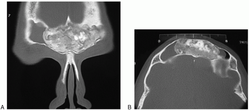 FIGURE 36.1 In coronal CT (A) and axial CT (B), the frontal osteoma completely fills the left frontal sinus and obstructs the outflow tract on the right side. Note how the osteoma appears to be pressed into the sinus conforming to the sinus-like modeling clay. |
PHYSICAL EXAMINATION
Mucoceles and tumors that erode the anterior table of the frontal sinus may present either with a localized swelling of the forehead or with proptosis. Tumors within the confines of the frontal sinus will often have few physical signs on examination. Nasal endoscopy is typically normal unless the condition is part of a chronic sinonasal disease such as chronic sinusitis. Septal deflections are noted and dealt with at the time of surgery. If the patient is bald, a discussion should be held with the patient about placement of the scalp incision. I prefer not to use eyebrow incisions as I think that even in bald patients, a coronal incision has a long-term better cosmetic result.
INDICATIONS
Large tumors (especially osteomas) that fill the frontal sinuses
Localized osteitis, especially laterally based
Lateral mucoceles
Lateral CSF leaks
Fractures of the frontal sinus
CONTRAINDICATIONS
There are few specific contraindications to OPF as an access procedure to the frontal sinuses.
Poorly pneumatized frontal sinuses make placing the osteotomies difficult as the sinuses are relatively small. These patients are often more suited to a Draf 3/modified Lothrop than an OPF.
Obliteration rather than a Draf 3 may be considered if the tumor involves the frontal sinus ostia and there is a narrow anteroposterior diameter and tumor removal entails removal of all mucosa in the region of the neo-ostium.
PREOPERATIVE PLANNING
Imaging Studies
Imaging studies should have been done as part of the diagnostic evaluation, and if they are done using the computerized image guidance protocols, then they can be used during surgery. If a tumor is present, then both CT and MRI scans need to be done. These are often merged so that the relationship between the soft tissue of the tumor and the bone of the frontal sinus can be studied. This will determine the best approach to the tumor. Image guidance is the preferred means to map the outline of the frontal sinuses on the anterior table of the frontal sinus before frontal sinus osteotomies are performed. Standard scuba strap or facial mask reference electrode/optical frame cannot be used for an OPF as these typically sit on the forehead in the region of the surgery. My preference is to place the head in a 3-pin brace with the electrode/optical frame attached to the brace. This prevents any obstruction of the image guidance reference to the frontal region.


